2HRV

2A CYSTEINE PROTEINASE FROM HUMAN RHINOVIRUS 2


Experimental Data Snapshot

  • Method: X-RAY DIFFRACTION
  • Resolution: 1.95 Å
  • R-Value Free: 0.250 
  • R-Value Work: 0.218 
  • R-Value Observed: 0.218 

wwPDB Validation   3D Report Full Report


This is version 1.3 of the entry. See complete history


Literature

The structure of the 2A proteinase from a common cold virus: a proteinase responsible for the shut-off of host-cell protein synthesis.

Petersen, J.F.Cherney, M.M.Liebig, H.D.Skern, T.Kuechler, E.James, M.N.

(1999) EMBO J 18: 5463-5475

  • DOI: https://doi.org/10.1093/emboj/18.20.5463
  • Primary Citation of Related Structures:  
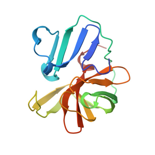
    2HRV

  • PubMed Abstract: 

    The crystal structure of the 2A proteinase from human rhinovirus serotype 2 (HRV2-2A(pro)) has been solved to 1.95 A resolution. The structure has an unusual, although chymotrypsin-related, fold comprising a unique four-stranded beta sheet as the N-terminal domain and a six-stranded beta barrel as the C-terminal domain. A tightly bound zinc ion, essential for the stability of HRV2-2A(pro), is tetrahedrally coordinated by three cysteine sulfurs and one histidine nitrogen. The active site consists of a catalytic triad formed by His18, Asp35 and Cys106. Asp35 is additionally involved in an extensive hydrogen-bonding network. Modelling studies reveal a substrate-induced fit that explains the specificity of the subsites S4, S2, S1 and S1'. The structure of HRV2-2A(pro) suggests the mechanism of the cis cleavage and its release from the polyprotein.


  • Organizational Affiliation

    MRC Group in Protein Structure and Function, Department of Biochemistry, University of Alberta, Edmonton, AB, Canada T6G 2H7.


Macromolecules
Find similar proteins by:  (by identity cutoff)  |  3D Structure
Entity ID: 1
MoleculeChains Sequence LengthOrganismDetailsImage
2A CYSTEINE PROTEINASE
A, B
142rhinovirus A2Mutation(s): 0 
EC: 3.4.22.29
UniProt
Find proteins for P04936 (Human rhinovirus 2)
Explore P04936 
Go to UniProtKB:  P04936
Entity Groups  
Sequence Clusters30% Identity50% Identity70% Identity90% Identity95% Identity100% Identity
UniProt GroupP04936
Sequence Annotations
Expand
  • Reference Sequence
Small Molecules
Ligands 1 Unique
IDChains Name / Formula / InChI Key2D Diagram3D Interactions
ZN
Query on ZN

Download Ideal Coordinates CCD File 
C [auth A],
D [auth B]
ZINC ION
Zn
PTFCDOFLOPIGGS-UHFFFAOYSA-N
Experimental Data & Validation

Experimental Data

  • Method: X-RAY DIFFRACTION
  • Resolution: 1.95 Å
  • R-Value Free: 0.250 
  • R-Value Work: 0.218 
  • R-Value Observed: 0.218 
  • Space Group: P 32 2 1
Unit Cell:
Length ( Å )Angle ( ˚ )
a = 72.921α = 90
b = 72.921β = 90
c = 161.659γ = 120
Software Package:
Software NamePurpose
SHELXSphasing
CCP4model building
DMmodel building
CNSrefinement
DENZOdata reduction
SCALEPACKdata scaling
CCP4phasing
DMphasing

Structure Validation

View Full Validation Report


Entry History 

Deposition Data

Revision History  (Full details and data files)

  • Version 1.0: 2000-05-03
    Type: Initial release
  • Version 1.1: 2008-04-26
    Changes: Version format compliance
  • Version 1.2: 2011-07-13
    Changes: Version format compliance
  • Version 1.3: 2023-12-27
    Changes: Data collection, Database references, Derived calculations