1RUC

RHINOVIRUS 14 MUTANT N1105S COMPLEXED WITH ANTIVIRAL COMPOUND WIN 52035


Experimental Data Snapshot

  • Method: X-RAY DIFFRACTION
  • Resolution: 3.10 Å

wwPDB Validation   3D Report Full Report


This is version 2.0 of the entry. See complete history


Literature

Structural studies on human rhinovirus 14 drug-resistant compensation mutants.

Hadfield, A.T.Oliveira, M.A.Kim, K.H.Minor, I.Kremer, M.J.Heinz, B.A.Shepard, D.Pevear, D.C.Rueckert, R.R.Rossmann, M.G.

(1995) J Mol Biol 253: 61-73

  • DOI: https://doi.org/10.1006/jmbi.1995.0536
  • Primary Citation of Related Structures:  
    1RUC, 1RUD, 1RUE, 1RUF, 1RUG, 1RUH, 1RUI, 1RUJ

  • PubMed Abstract: 

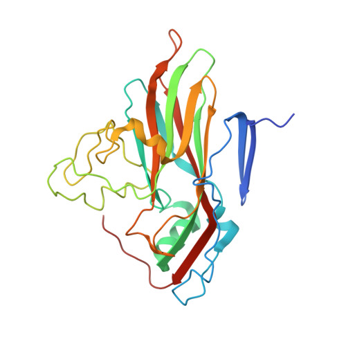
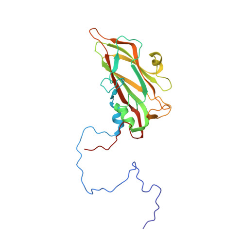
    Structures have been determined of three human rhinovirus 14 (HRV14) compensation mutants that have resistance to the antiviral capsid binding compounds WIN 52035 and WIN 52084. In addition, the structure of HRV14 is reported, with a site-directed mutation at residue 1219 in VP1. A spontaneous mutation occurs at the same site in one of the compensation mutants. Some of the mutations are on the viral surface in the canyon and some lie within the hydrophobic binding pocket in VP1 below the ICAM footprint. Those mutant virus strains with mutations on the surface bind better to cells than does wild-type virus. The antiviral compounds bind to the mutant viruses in a manner similar to their binding to wild-type virus. The receptor and WIN compound binding sites overlap, causing competition between receptor attachment and antiviral compound binding. The compensation mutants probably function by shifting the equilibrium in favor of receptor binding. The mutations in the canyon increase the affinity of the virus for the receptor, while the mutations in the pocket probably decrease the affinity of the WIN compounds for the virus by reducing favorable hydrophobic contacts and constricting the pore through which the antiviral compounds are thought to enter the pocket. This is in contrast to the resistant exclusion mutants that block compounds from binding by increasing the bulk of residues within the hydrophobic pocket in VP1.


  • Organizational Affiliation

    Department of Biological Sciences, Purdue University, West Lafayette, IN 47907-1392, USA.


Macromolecules
Find similar proteins by:  (by identity cutoff)  |  3D Structure
Entity ID: 1
MoleculeChains Sequence LengthOrganismDetailsImage
RHINOVIRUS 14A [auth 1]289rhinovirus B14Mutation(s): 1 
UniProt
Find proteins for P03303 (Human rhinovirus 14)
Explore P03303 
Go to UniProtKB:  P03303
Entity Groups  
Sequence Clusters30% Identity50% Identity70% Identity90% Identity95% Identity100% Identity
UniProt GroupP03303
Sequence Annotations
Expand
  • Reference Sequence
Find similar proteins by:  (by identity cutoff)  |  3D Structure
Entity ID: 2
MoleculeChains Sequence LengthOrganismDetailsImage
RHINOVIRUS 14B [auth 2]262rhinovirus B14Mutation(s): 0 
UniProt
Find proteins for P03303 (Human rhinovirus 14)
Explore P03303 
Go to UniProtKB:  P03303
Entity Groups  
Sequence Clusters30% Identity50% Identity70% Identity90% Identity95% Identity100% Identity
UniProt GroupP03303
Sequence Annotations
Expand
  • Reference Sequence
Find similar proteins by:  (by identity cutoff)  |  3D Structure
Entity ID: 3
MoleculeChains Sequence LengthOrganismDetailsImage
RHINOVIRUS 14C [auth 3]236rhinovirus B14Mutation(s): 0 
UniProt
Find proteins for P03303 (Human rhinovirus 14)
Explore P03303 
Go to UniProtKB:  P03303
Entity Groups  
Sequence Clusters30% Identity50% Identity70% Identity90% Identity95% Identity100% Identity
UniProt GroupP03303
Sequence Annotations
Expand
  • Reference Sequence
Find similar proteins by:  (by identity cutoff)  |  3D Structure
Entity ID: 4
MoleculeChains Sequence LengthOrganismDetailsImage
RHINOVIRUS 14D [auth 4]68rhinovirus B14Mutation(s): 0 
UniProt
Find proteins for P03303 (Human rhinovirus 14)
Explore P03303 
Go to UniProtKB:  P03303
Entity Groups  
Sequence Clusters30% Identity50% Identity70% Identity90% Identity95% Identity100% Identity
UniProt GroupP03303
Sequence Annotations
Expand
  • Reference Sequence
Small Molecules
Ligands 1 Unique
IDChains Name / Formula / InChI Key2D Diagram3D Interactions
W35
Query on W35

Download Ideal Coordinates CCD File 
E [auth 1]5-(5-(4-(4,5-DIHYDRO-2-OXAZOLY)PHENOXY)PENTYL)-3-METHYL ISOXAZOLE
C18 H22 N2 O3
IWZDYGHUSXWPPM-UHFFFAOYSA-N
Experimental Data & Validation

Experimental Data

  • Method: X-RAY DIFFRACTION
  • Resolution: 3.10 Å
  • Space Group: P 21 3
Unit Cell:
Length ( Å )Angle ( ˚ )
a = 445.1α = 90
b = 445.1β = 90
c = 445.1γ = 90
Software Package:
Software NamePurpose
Omodel building
PURDUEdata reduction

Structure Validation

View Full Validation Report


Entry History 

Deposition Data

Revision History  (Full details and data files)

  • Version 1.0: 1995-11-14
    Type: Initial release
  • Version 1.1: 2008-03-24
    Changes: Version format compliance
  • Version 1.2: 2011-07-13
    Changes: Version format compliance
  • Version 1.3: 2021-11-03
    Changes: Data collection, Database references, Derived calculations
  • Version 2.0: 2023-01-18
    Type: Remediation
    Changes: Advisory, Atomic model, Data collection, Derived calculations, Other, Refinement description