5V0L

Crystal structure of the AHR-ARNT heterodimer in complex with the DRE


Experimental Data Snapshot

  • Method: X-RAY DIFFRACTION
  • Resolution: 4.00 Å
  • R-Value Free: 0.323 
  • R-Value Work: 0.285 
  • R-Value Observed: 0.287 

wwPDB Validation   3D Report Full Report


This is version 1.5 of the entry. See complete history


Literature

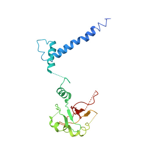
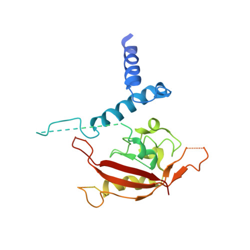
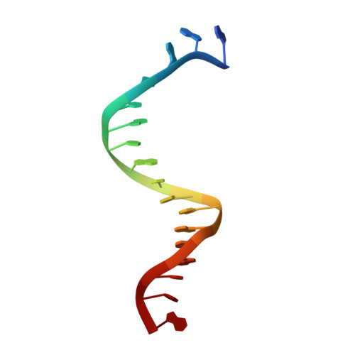
Structural hierarchy controlling dimerization and target DNA recognition in the AHR transcriptional complex.

Seok, S.H.Lee, W.Jiang, L.Molugu, K.Zheng, A.Li, Y.Park, S.Bradfield, C.A.Xing, Y.

(2017) Proc Natl Acad Sci U S A 114: 5431-5436

  • DOI: https://doi.org/10.1073/pnas.1617035114
  • Primary Citation of Related Structures:  
    5V0L

  • PubMed Abstract: 

    The aryl hydrocarbon receptor (AHR) belongs to the PAS (PER-ARNT-SIM) family transcription factors and mediates broad responses to numerous environmental pollutants and cellular metabolites, modulating diverse biological processes from adaptive metabolism, acute toxicity, to normal physiology of vascular and immune systems. The AHR forms a transcriptionally active heterodimer with ARNT (AHR nuclear translocator), which recognizes the dioxin response element (DRE) in the promoter of downstream genes. We determined the crystal structure of the mammalian AHR-ARNT heterodimer in complex with the DRE, in which ARNT curls around AHR into a highly intertwined asymmetric architecture, with extensive heterodimerization interfaces and AHR interdomain interactions. Specific recognition of the DRE is determined locally by the DNA-binding residues, which discriminates it from the closely related hypoxia response element (HRE), and is globally affected by the dimerization interfaces and interdomain interactions. Changes at the interdomain interactions caused either AHR constitutive nuclear localization or failure to translocate to nucleus, underlying an allosteric structural pathway for mediating ligand-induced exposure of nuclear localization signal. These observations, together with the global higher flexibility of the AHR PAS-A and its loosely packed structural elements, suggest a dynamic structural hierarchy for complex scenarios of AHR activation induced by its diverse ligands.


  • Organizational Affiliation

    McArdle Laboratory for Cancer Research, Department of Oncology, School of Medicine and Public Health, University of Wisconsin, Madison, WI 53705.


Macromolecules

Find similar proteins by:  (by identity cutoff)  |  3D Structure
Entity ID: 1
MoleculeChains Sequence LengthOrganismDetailsImage
Aryl hydrocarbon receptor nuclear translocator279Homo sapiensMutation(s): 0 
Gene Names: ARNTBHLHE2
UniProt & NIH Common Fund Data Resources
Find proteins for P27540 (Homo sapiens)
Explore P27540 
Go to UniProtKB:  P27540
PHAROS:  P27540
GTEx:  ENSG00000143437 
Entity Groups  
Sequence Clusters30% Identity50% Identity70% Identity90% Identity95% Identity100% Identity
UniProt GroupP27540
Sequence Annotations
Expand
  • Reference Sequence
Find similar proteins by:  (by identity cutoff)  |  3D Structure
Entity ID: 2
MoleculeChains Sequence LengthOrganismDetailsImage
Aryl hydrocarbon receptor241Mus musculusMutation(s): 0 
Gene Names: Ahr
UniProt
Find proteins for P30561 (Mus musculus)
Explore P30561 
Go to UniProtKB:  P30561
Entity Groups  
Sequence Clusters30% Identity50% Identity70% Identity90% Identity95% Identity100% Identity
UniProt GroupP30561
Sequence Annotations
Expand
  • Reference Sequence

Find similar nucleic acids by:  Sequence   |   3D Structure  

Entity ID: 3
MoleculeChains LengthOrganismImage
DNA (5'-D(P*GP*GP*AP*TP*TP*GP*CP*GP*TP*GP*AP*GP*AP*AP*CP*TP*G)-3')17Homo sapiens
Sequence Annotations
Expand
  • Reference Sequence

Find similar nucleic acids by:  Sequence   |   3D Structure  

Entity ID: 4
MoleculeChains LengthOrganismImage
DNA (5'-D(P*AP*GP*TP*TP*CP*TP*CP*AP*CP*GP*CP*AP*AP*T)-3')14Homo sapiens
Sequence Annotations
Expand
  • Reference Sequence
Experimental Data & Validation

Experimental Data

  • Method: X-RAY DIFFRACTION
  • Resolution: 4.00 Å
  • R-Value Free: 0.323 
  • R-Value Work: 0.285 
  • R-Value Observed: 0.287 
  • Space Group: C 1 2 1
Unit Cell:
Length ( Å )Angle ( ˚ )
a = 78.199α = 90
b = 64.364β = 100.14
c = 157.725γ = 90
Software Package:
Software NamePurpose
PHENIXrefinement
HKL-2000data scaling
PHENIXphasing
HKL-2000data reduction

Structure Validation

View Full Validation Report


Entry History & Funding Information

Deposition Data


Funding OrganizationLocationGrant Number
National Institutes of Health/National Institute of General Medical Sciences (NIH/NIGMS)United StatesR01 GM096060-01

Revision History  (Full details and data files)

  • Version 1.0: 2017-04-19
    Type: Initial release
  • Version 1.1: 2017-04-26
    Changes: Database references
  • Version 1.2: 2017-06-07
    Changes: Database references
  • Version 1.3: 2017-09-27
    Changes: Author supporting evidence
  • Version 1.4: 2020-01-01
    Changes: Author supporting evidence
  • Version 1.5: 2023-10-04
    Changes: Data collection, Database references, Refinement description