1W9O

Crystal structure of the PDZ tandem of human syntenin in complex with TNEYYV peptide


Experimental Data Snapshot

  • Method: X-RAY DIFFRACTION
  • Resolution: 2.25 Å
  • R-Value Free: 0.281 
  • R-Value Work: 0.214 
  • R-Value Observed: 0.218 

wwPDB Validation   3D Report Full Report


This is version 1.3 of the entry. See complete history


Literature

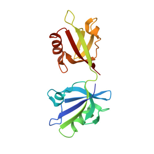
The Binding of the Pdz Tandem of Syntenin to Target Proteins.

Grembecka, J.Cierpicki, T.Devedjiev, Y.Derewenda, U.Kang, B.S.Bushweller, J.H.Derewenda, Z.S.

(2006) Biochemistry 45: 3674

  • DOI: https://doi.org/10.1021/bi052225y
  • Primary Citation of Related Structures:  
    1V1T, 1W9E, 1W9O, 1W9Q, 1YBO

  • PubMed Abstract: 

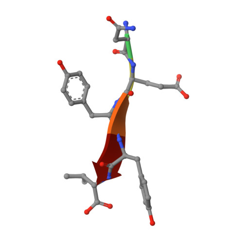
    PDZ domains are among the most abundant protein modules in the known genomes. Their main function is to provide scaffolds for membrane-associated protein complexes by binding to the cytosolic, C-terminal fragments of receptors, channels, and other integral membrane proteins. Here, using both heteronuclear NMR and single crystal X-ray diffraction, we show how peptides with different sequences, including those corresponding to the C-termini of syndecan, neurexin, and ephrin B, can simultaneously bind to both PDZ domains of the scaffolding protein syntenin. The PDZ2 domain binds these peptides in the canonical fashion, and an induced fit mechanism allows for the accommodation of a range of side chains in the P(0) and P(-)(2) positions. However, binding to the PDZ1 domain requires that the target peptide assume a noncanonical conformation. These data help explain how syntenin, and perhaps other PDZ-containing proteins, may preferentially bind to dimeric and clustered targets, and provide a mechanistic explanation for the previously reported cooperative ligand binding by syntenin's two PDZ domains.


  • Organizational Affiliation

    Department of Molecular Physiology and Biological Physics, University of Virginia School of Medicine, Charlottesville, Virginia 22908-0736, USA.


Macromolecules
Find similar proteins by:  (by identity cutoff)  |  3D Structure
Entity ID: 1
MoleculeChains Sequence LengthOrganismDetailsImage
SYNTENIN 1
A, B
166Homo sapiensMutation(s): 0 
Gene Names: SDCBPMDA9SYCL
UniProt & NIH Common Fund Data Resources
Find proteins for O00560 (Homo sapiens)
Explore O00560 
Go to UniProtKB:  O00560
PHAROS:  O00560
GTEx:  ENSG00000137575 
Entity Groups  
Sequence Clusters30% Identity50% Identity70% Identity90% Identity95% Identity100% Identity
UniProt GroupO00560
Sequence Annotations
Expand
  • Reference Sequence

Find similar proteins by:  Sequence   |   3D Structure  

Entity ID: 2
MoleculeChains Sequence LengthOrganismDetailsImage
TNEYYV PEPTIDEC [auth S],
D [auth T]
6synthetic constructMutation(s): 0 
Sequence Annotations
Expand
  • Reference Sequence
Small Molecules
Ligands 1 Unique
IDChains Name / Formula / InChI Key2D Diagram3D Interactions
BEZ
Query on BEZ

Download Ideal Coordinates CCD File 
E [auth A]BENZOIC ACID
C7 H6 O2
WPYMKLBDIGXBTP-UHFFFAOYSA-N
Experimental Data & Validation

Experimental Data

  • Method: X-RAY DIFFRACTION
  • Resolution: 2.25 Å
  • R-Value Free: 0.281 
  • R-Value Work: 0.214 
  • R-Value Observed: 0.218 
  • Space Group: P 41 21 2
Unit Cell:
Length ( Å )Angle ( ˚ )
a = 72.134α = 90
b = 72.134β = 90
c = 126.221γ = 90
Software Package:
Software NamePurpose
REFMACrefinement
HKL-2000data reduction
HKL-2000data scaling
AMoREphasing

Structure Validation

View Full Validation Report


Entry History 

Deposition Data

Revision History  (Full details and data files)

  • Version 1.0: 2006-03-22
    Type: Initial release
  • Version 1.1: 2012-05-30
    Changes: Derived calculations, Non-polymer description, Other, Structure summary, Version format compliance
  • Version 1.2: 2019-01-30
    Changes: Data collection, Experimental preparation, Source and taxonomy
  • Version 1.3: 2023-12-13
    Changes: Data collection, Database references, Derived calculations, Other, Refinement description