Structure of the Ubiquitin-associated Domain of p62 (SQSTM1) and Implications for Mutations That Cause Paget's Disease of Bone
Ciani, B., Layfield, R., Cavey, J.R., Sheppard, P.W., Searle, M.S.(2003) J Biol Chem 278: 37409-37412
- PubMed: 12857745
- DOI: https://doi.org/10.1074/jbc.M307416200
- Primary Citation of Related Structures:
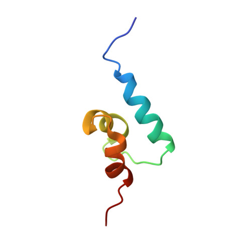
1Q02 - PubMed Abstract:
The p62 protein (also known as SQSTM1) mediates diverse cellular functions including control of NFkappaB signaling and transcriptional activation. p62 binds non-covalently to ubiquitin and co-localizes with ubiquitylated inclusions in a number of human protein aggregation diseases. Mutations in the gene encoding p62 cause Paget's disease of bone (PDB), a common disorder of the elderly characterized by excessive bone resorption and formation. All of the p62 PDB mutations identified to date cluster within the C-terminal region of the protein, which shows low sequence identity to previously characterized ubiquitin-associated (UBA) domains. We report the first NMR structure of a recombinant polypeptide that contains the C-terminal UBA domain of the human p62 protein (residues 387-436). This sequence, which confers multiubiquitin chain binding, forms a compact three-helix bundle with a structure analogous to the UBA domains of HHR23A but with differences in the loop regions connecting helices that may be involved in binding accessory proteins. We show that the Pro392 --> Leu PDB substitution mutation modifies the structure of the UBA domain by extending the N terminus of helix 1. In contrast to the p62 PDB deletion mutations that remove the UBA domain and ablate multiubiquitin chain binding, the Pro392 --> Leu substitution does not affect interaction of the UBA domain with multiubiquitin chains. Thus, phenotypically identical substitution and deletion mutations do not appear to predispose to PDB through a mechanism dependent on a common loss of ubiquitin chain binding by p62.
Organizational Affiliation:
School of Chemistry, University Park, Nottingham NG7 2RD, United Kingdom.