8DML

Vibrio parahaemolyticus VtrA/VtrC complex bound to the bile salt chenodeoxycholate


Experimental Data Snapshot

  • Method: X-RAY DIFFRACTION
  • Resolution: 2.08 Å
  • R-Value Free: 0.224 
  • R-Value Work: 0.188 
  • R-Value Observed: 0.190 

wwPDB Validation   3D Report Full Report


Ligand Structure Quality Assessment 


This is version 1.1 of the entry. See complete history


Literature

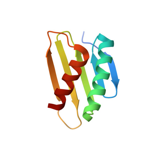
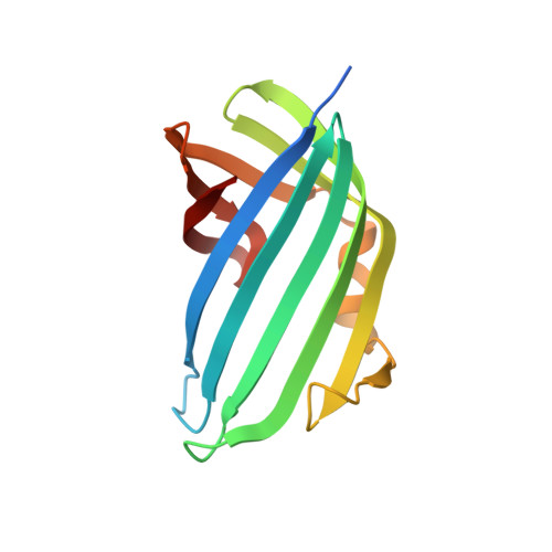
Molecular determinants for differential activation of the bile acid receptor from the pathogen Vibrio parahaemolyticus.

Zou, A.J.Kinch, L.Chimalapati, S.Garcia, N.Tomchick, D.R.Orth, K.

(2023) J Biol Chem 299: 104591-104591

  • DOI: https://doi.org/10.1016/j.jbc.2023.104591
  • Primary Citation of Related Structures:  
    8DML

  • PubMed Abstract: 

    Bile acids are important for digestion of food and antimicrobial activity. Pathogenic Vibrio parahaemolyticus senses bile acids and induce pathogenesis. The bile acid taurodeoxycholate (TDC) was shown to activate the master regulator, VtrB, of this system, whereas other bile acids such as chenodeoxycholate (CDC) do not. Previously, VtrA-VtrC was discovered to be the co-component signal transduction system that binds bile acids and induces pathogenesis. TDC binds to the periplasmic domain of the VtrA-VtrC complex, activating a DNA-binding domain in VtrA that then activates VtrB. Here, we find that CDC and TDC compete for binding to the VtrA-VtrC periplasmic heterodimer. Our crystal structure of the VtrA-VtrC heterodimer bound to CDC revealed CDC binds in the same hydrophobic pocket as TDC but differently. Using isothermal titration calorimetry, we observed that most mutants in the binding pocket of VtrA-VtrC caused a decrease in bile acid binding affinity. Notably, two mutants in VtrC bound bile acids with a similar affinity as the WT protein but were attenuated for TDC-induced type III secretion system 2 activation. Collectively, these studies provide a molecular explanation for the selective pathogenic signaling by V. parahaemolyticus and reveal insight into a host's susceptibility to disease.


  • Organizational Affiliation

    Department of Molecular Biology, University of Texas Southwestern Medical Center, Dallas, Texas, USA.


Macromolecules
Find similar proteins by:  (by identity cutoff)  |  3D Structure
Entity ID: 1
MoleculeChains Sequence LengthOrganismDetailsImage
VtrA
A, C, E, G
94Vibrio parahaemolyticusMutation(s): 0 
Gene Names: VPA1332
UniProt
Find proteins for Q87GI4 (Vibrio parahaemolyticus serotype O3:K6 (strain RIMD 2210633))
Explore Q87GI4 
Go to UniProtKB:  Q87GI4
Entity Groups  
Sequence Clusters30% Identity50% Identity70% Identity90% Identity95% Identity100% Identity
UniProt GroupQ87GI4
Sequence Annotations
Expand
  • Reference Sequence
Find similar proteins by:  (by identity cutoff)  |  3D Structure
Entity ID: 2
MoleculeChains Sequence LengthOrganismDetailsImage
VtrC
B, D, F, H
144Vibrio parahaemolyticusMutation(s): 0 
Gene Names: VPA1333
UniProt
Find proteins for Q87GI3 (Vibrio parahaemolyticus serotype O3:K6 (strain RIMD 2210633))
Explore Q87GI3 
Go to UniProtKB:  Q87GI3
Entity Groups  
Sequence Clusters30% Identity50% Identity70% Identity90% Identity95% Identity100% Identity
UniProt GroupQ87GI3
Sequence Annotations
Expand
  • Reference Sequence
Small Molecules
Ligands 4 Unique
IDChains Name / Formula / InChI Key2D Diagram3D Interactions
JN3 (Subject of Investigation/LOI)
Query on JN3

Download Ideal Coordinates CCD File 
J [auth B],
Q [auth D],
V [auth F],
Y [auth H]
CHENODEOXYCHOLIC ACID
C24 H40 O4
RUDATBOHQWOJDD-BSWAIDMHSA-N
PG4
Query on PG4

Download Ideal Coordinates CCD File 
L [auth B],
S [auth D],
W [auth F],
Z [auth H]
TETRAETHYLENE GLYCOL
C8 H18 O5
UWHCKJMYHZGTIT-UHFFFAOYSA-N
EDO
Query on EDO

Download Ideal Coordinates CCD File 
BA [auth H]
I [auth A]
K [auth B]
N [auth B]
O [auth B]
BA [auth H],
I [auth A],
K [auth B],
N [auth B],
O [auth B],
P [auth C],
R [auth D],
U [auth D]
1,2-ETHANEDIOL
C2 H6 O2
LYCAIKOWRPUZTN-UHFFFAOYSA-N
CA
Query on CA

Download Ideal Coordinates CCD File 
AA [auth H],
M [auth B],
T [auth D],
X [auth F]
CALCIUM ION
Ca
BHPQYMZQTOCNFJ-UHFFFAOYSA-N
Experimental Data & Validation

Experimental Data

  • Method: X-RAY DIFFRACTION
  • Resolution: 2.08 Å
  • R-Value Free: 0.224 
  • R-Value Work: 0.188 
  • R-Value Observed: 0.190 
  • Space Group: C 1 2 1
Unit Cell:
Length ( Å )Angle ( ˚ )
a = 142.603α = 90
b = 41.763β = 91.57
c = 168.956γ = 90
Software Package:
Software NamePurpose
HKL-3000data scaling
PHENIXrefinement
PDB_EXTRACTdata extraction
HKL-3000data reduction
PHENIXphasing

Structure Validation

View Full Validation Report


Ligand Structure Quality Assessment 


Entry History & Funding Information

Deposition Data


Funding OrganizationLocationGrant Number
National Institutes of Health/National Institute of General Medical Sciences (NIH/NIGMS)United States130305
Robert A. Welch FoundationUnited StatesI-1561
National Institutes of Health/National Institute of General Medical Sciences (NIH/NIGMS)United StatesS10OD025018

Revision History  (Full details and data files)

  • Version 1.0: 2023-06-14
    Type: Initial release
  • Version 1.1: 2023-10-25
    Changes: Data collection, Refinement description