5EUH

Crystal structure of the c-di-GMP-bound GGDEF domain of P. fluorescens GcbC

  • Classification: MEMBRANE PROTEIN
  • Organism(s): Pseudomonas fluorescens Pf0-1
  • Expression System: Escherichia coli
  • Mutation(s): No 

  • Deposited: 2015-11-18 Released: 2015-12-30 
  • Deposition Author(s): Giglio, K.M., Cooley, R.B., Sondermann, H.
  • Funding Organization(s): National Institutes of Health/National Institute Of Allergy and Infectious Diseases (NIH/NIAID), National Institutes of Health/National Institute of General Medical Sciences (NIH/NIGMS), National Science Foundation (NSF, United States)

Experimental Data Snapshot

  • Method: X-RAY DIFFRACTION
  • Resolution: 2.99 Å
  • R-Value Free: 0.243 
  • R-Value Work: 0.226 
  • R-Value Observed: 0.227 

wwPDB Validation   3D Report Full Report


Ligand Structure Quality Assessment 


This is version 1.4 of the entry. See complete history


Literature

Contribution of Physical Interactions to Signaling Specificity between a Diguanylate Cyclase and Its Effector.

Dahlstrom, K.M.Giglio, K.M.Collins, A.J.Sondermann, H.O'Toole, G.A.

(2015) mBio 6: e01978-e01915

  • DOI: https://doi.org/10.1128/mBio.01978-15
  • Primary Citation of Related Structures:  
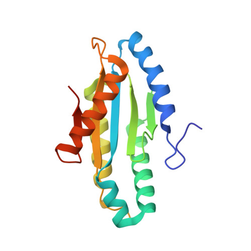
    5EUH

  • PubMed Abstract: 

    Cyclic diguanylate (c-di-GMP) is a bacterial second messenger that controls multiple cellular processes. c-di-GMP networks have up to dozens of diguanylate cyclases (DGCs) that synthesize c-di-GMP along with many c-di-GMP-responsive target proteins that can bind and respond to this signal. For such networks to have order, a mechanism(s) likely exists that allow DGCs to specifically signal their targets, and it has been suggested that physical interactions might provide such specificity. Our results show a DGC from Pseudomonas fluorescens physically interacting with its target protein at a conserved interface, and this interface can be predictive of DGC-target protein interactions. Furthermore, we demonstrate that physical interaction is necessary for the DGC to maximally signal its target. If such "local signaling" is a theme for even a fraction of the DGCs used by bacteria, it becomes possible to posit a model whereby physical interaction allows a DGC to directly signal its target protein, which in turn may help curtail undesired cross talk with other members of the network.


  • Organizational Affiliation

    Department of Microbiology and Immunology, Geisel School of Medicine at Dartmouth, Hanover, New Hampshire.


Macromolecules
Find similar proteins by:  (by identity cutoff)  |  3D Structure
Entity ID: 1
MoleculeChains Sequence LengthOrganismDetailsImage
Putative GGDEF domain membrane protein
A, B, C, D
172Pseudomonas fluorescens Pf0-1Mutation(s): 0 
Gene Names: Pfl01_4666
UniProt
Find proteins for Q3K751 (Pseudomonas fluorescens (strain Pf0-1))
Explore Q3K751 
Go to UniProtKB:  Q3K751
Entity Groups  
Sequence Clusters30% Identity50% Identity70% Identity90% Identity95% Identity100% Identity
UniProt GroupQ3K751
Sequence Annotations
Expand
  • Reference Sequence
Small Molecules
Ligands 2 Unique
IDChains Name / Formula / InChI Key2D Diagram3D Interactions
C2E
Query on C2E

Download Ideal Coordinates CCD File 
E [auth A],
H [auth B],
I [auth B],
M [auth C]
9,9'-[(2R,3R,3aS,5S,7aR,9R,10R,10aS,12S,14aR)-3,5,10,12-tetrahydroxy-5,12-dioxidooctahydro-2H,7H-difuro[3,2-d:3',2'-j][1,3,7,9,2,8]tetraoxadiphosphacyclododecine-2,9-diyl]bis(2-amino-1,9-dihydro-6H-purin-6-one)
C20 H24 N10 O14 P2
PKFDLKSEZWEFGL-MHARETSRSA-N
SO4
Query on SO4

Download Ideal Coordinates CCD File 
F [auth A],
G [auth A],
J [auth B],
K [auth B],
L [auth B]
SULFATE ION
O4 S
QAOWNCQODCNURD-UHFFFAOYSA-L
Experimental Data & Validation

Experimental Data

  • Method: X-RAY DIFFRACTION
  • Resolution: 2.99 Å
  • R-Value Free: 0.243 
  • R-Value Work: 0.226 
  • R-Value Observed: 0.227 
  • Space Group: P 62
Unit Cell:
Length ( Å )Angle ( ˚ )
a = 141.913α = 90
b = 141.913β = 90
c = 106.169γ = 120
Software Package:
Software NamePurpose
PHENIXrefinement
HKL-2000data reduction
HKL-2000data scaling
PHENIXphasing

Structure Validation

View Full Validation Report


Ligand Structure Quality Assessment 


Entry History & Funding Information

Deposition Data


Funding OrganizationLocationGrant Number
National Institutes of Health/National Institute Of Allergy and Infectious Diseases (NIH/NIAID)United StatesR01-AI097307
National Institutes of Health/National Institute of General Medical Sciences (NIH/NIGMS)United StatesGM-103485
National Science Foundation (NSF, United States)United StatesDMR-1332208

Revision History  (Full details and data files)

  • Version 1.0: 2015-12-30
    Type: Initial release
  • Version 1.1: 2016-02-10
    Changes: Database references
  • Version 1.2: 2017-09-27
    Changes: Author supporting evidence, Derived calculations
  • Version 1.3: 2019-11-27
    Changes: Author supporting evidence
  • Version 1.4: 2023-09-27
    Changes: Data collection, Database references, Refinement description, Structure summary