5FLH

Free state of Ni-quercetinase


Experimental Data Snapshot

  • Method: X-RAY DIFFRACTION
  • Resolution: 1.80 Å
  • R-Value Free: 0.230 
  • R-Value Work: 0.189 
  • R-Value Observed: 0.191 

wwPDB Validation   3D Report Full Report


This is version 1.1 of the entry. See complete history


Literature

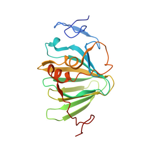
Quercetin 2,4-Dioxygenase Activates Dioxygen in a Side-on O2 -Ni Complex.

Jeoung, J.Nianios, D.Fetzner, S.Dobbek, H.

(2016) Angew Chem Int Ed Engl 55: 3281

  • DOI: https://doi.org/10.1002/anie.201510741
  • Primary Citation of Related Structures:  
    5FLH, 5FLI, 5FLJ

  • PubMed Abstract: 

    Quercetin 2,4-dioxygenase (quercetinase) from Streptomyces uses nickel as the active-site cofactor to catalyze oxidative cleavage of the flavonol quercetin. How this unusual active-site metal supports catalysis and O2 activation is under debate. We present crystal structures of Ni-quercetinase in three different states, thus providing direct insight into how quercetin and O2 are activated at the Ni(2+) ion. The Ni(2+) ion is coordinated by three histidine residues and a glutamate residue (E(76)) in all three states. Upon binding, quercetin replaces one water ligand at Ni and is stabilized by a short hydrogen bond through E(76) , the carboxylate group of which rotates by 90°. This conformational change weakens the interaction between Ni and the remaining water ligand, thereby preparing a coordination site at Ni to bind O2. O2 binds side-on to the Ni(2+) ion and is perpendicular to the C2-C3 and C3-C4 bonds of quercetin, which are cleaved in the following reaction steps.


  • Organizational Affiliation

    Institut für Biologie, Strukturbiologie/Biochemie, Humboldt-Universität zu Berlin, Unter den Linden 6, 10099, Berlin, Germany.


Macromolecules
Find similar proteins by:  (by identity cutoff)  |  3D Structure
Entity ID: 1
MoleculeChains Sequence LengthOrganismDetailsImage
QUERCETINASE QUED
A, B
192Streptomyces sp. FLAMutation(s): 0 
Gene Names: QUED
UniProt
Find proteins for A2VA43 (Streptomyces sp. FLA)
Explore A2VA43 
Go to UniProtKB:  A2VA43
Entity Groups  
Sequence Clusters30% Identity50% Identity70% Identity90% Identity95% Identity100% Identity
UniProt GroupA2VA43
Sequence Annotations
Expand
  • Reference Sequence
Experimental Data & Validation

Experimental Data

  • Method: X-RAY DIFFRACTION
  • Resolution: 1.80 Å
  • R-Value Free: 0.230 
  • R-Value Work: 0.189 
  • R-Value Observed: 0.191 
  • Space Group: P 61 2 2
Unit Cell:
Length ( Å )Angle ( ˚ )
a = 107.692α = 90
b = 107.692β = 90
c = 144.067γ = 120
Software Package:
Software NamePurpose
PHENIXrefinement
XDSdata reduction
XSCALEdata scaling

Structure Validation

View Full Validation Report


Entry History 

Deposition Data

Revision History  (Full details and data files)

  • Version 1.0: 2016-06-01
    Type: Initial release
  • Version 1.1: 2017-08-23
    Changes: Data collection, Structure summary