3ESL

Crystal structure of the conserved N-terminal domain of the mitotic checkpoint component BUB1


Experimental Data Snapshot

  • Method: X-RAY DIFFRACTION
  • Resolution: 1.74 Å
  • R-Value Free: 0.216 
  • R-Value Work: 0.190 
  • R-Value Observed: 0.190 

wwPDB Validation   3D Report Full Report


This is version 1.2 of the entry. See complete history


Literature

The Crystal Structure of the N-Terminal Region of BUB1 Provides Insight into the Mechanism of BUB1 Recruitment to Kinetochores.

Bolanos-Garcia, V.M.Kiyomitsu, T.D'Arcy, S.Chirgadze, D.Y.Grossmann, J.G.Matak-Vinkovic, D.Venkitaraman, A.R.Yanagida, M.Robinson, C.V.Blundell, T.L.

(2009) Structure 17: 105-116

  • DOI: https://doi.org/10.1016/j.str.2008.10.015
  • Primary Citation of Related Structures:  
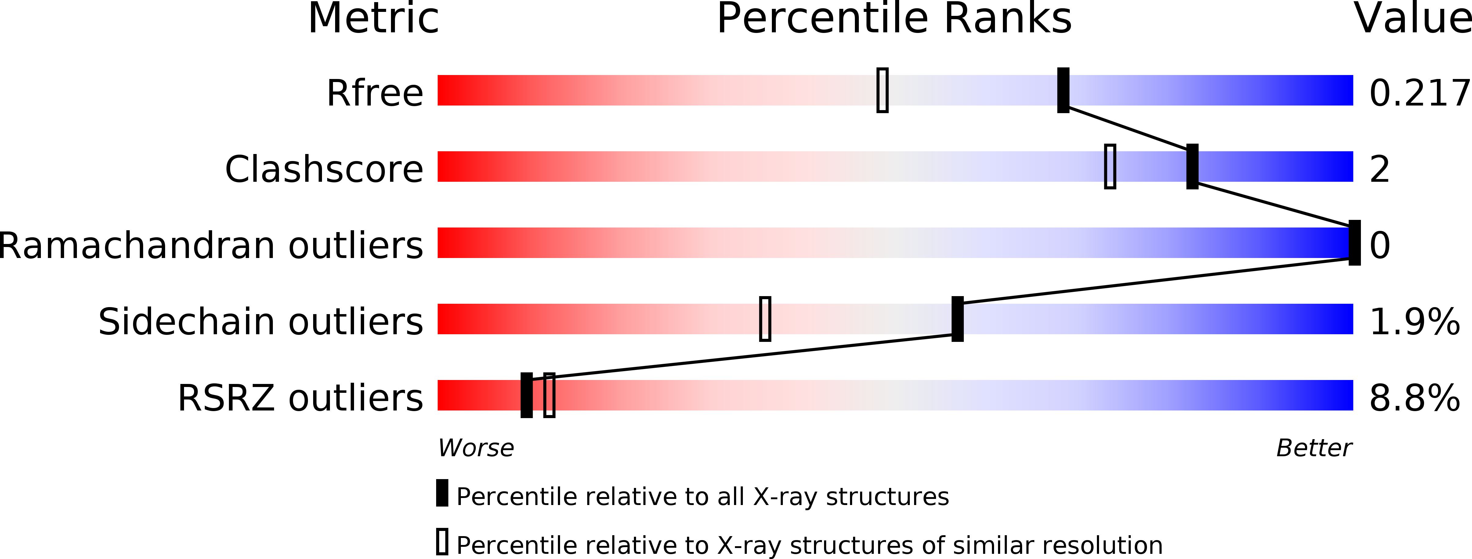
    3ESL

  • PubMed Abstract: 

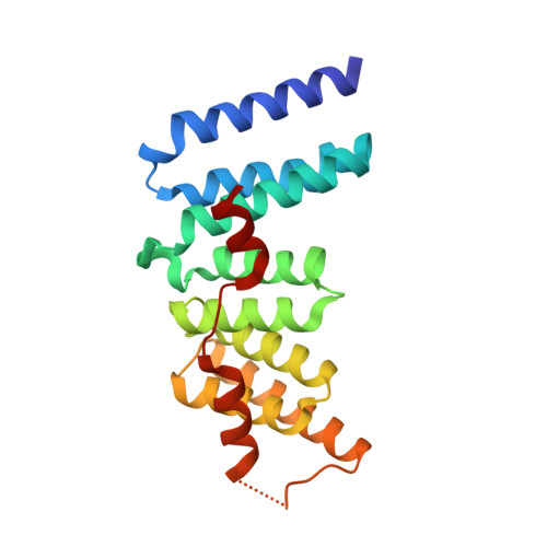
    The interaction of the central mitotic checkpoint component BUB1 with the mitotic kinetochore protein Blinkin is required for the kinetochore localization and function of BUB1 in the mitotic spindle assembly checkpoint, the regulatory mechanism of the cell cycle that ensures the even distribution of chromosomes during the transition from metaphase to anaphase. Here, we report the 1.74 angstroms resolution crystal structure of the N-terminal region of BUB1. The structure is organized as a tandem arrangement of three divergent units of the tetratricopeptide motif. Functional assays in vivo of native and site-specific mutants identify the residues of human BUB1 important for the interaction with Blinkin and define one region of potential therapeutic interest. The structure provides insight into the molecular basis of Blinkin-specific recognition by BUB1 and, on a broader perspective, of the mechanism that mediates kinetochore localization of BUB1 in checkpoint-activated cells.


  • Organizational Affiliation

    Department of Biochemistry, University of Cambridge, Cambridge CB2 1GA, UK. victor@cryst.bioc.cam.ac.uk


Macromolecules
Find similar proteins by:  (by identity cutoff)  |  3D Structure
Entity ID: 1
MoleculeChains Sequence LengthOrganismDetailsImage
Checkpoint serine/threonine-protein kinase BUB1
A, B
202Saccharomyces cerevisiaeMutation(s): 0 
Gene Names: BUB1G7542YGR188C
UniProt
Find proteins for A6ZUJ9 (Saccharomyces cerevisiae (strain YJM789))
Explore A6ZUJ9 
Go to UniProtKB:  A6ZUJ9
Entity Groups  
Sequence Clusters30% Identity50% Identity70% Identity90% Identity95% Identity100% Identity
UniProt GroupA6ZUJ9
Sequence Annotations
Expand
  • Reference Sequence
Small Molecules
Ligands 1 Unique
IDChains Name / Formula / InChI Key2D Diagram3D Interactions
NHE
Query on NHE

Download Ideal Coordinates CCD File 
C [auth A],
D [auth B]
2-[N-CYCLOHEXYLAMINO]ETHANE SULFONIC ACID
C8 H17 N O3 S
MKWKNSIESPFAQN-UHFFFAOYSA-N
Experimental Data & Validation

Experimental Data

  • Method: X-RAY DIFFRACTION
  • Resolution: 1.74 Å
  • R-Value Free: 0.216 
  • R-Value Work: 0.190 
  • R-Value Observed: 0.190 
  • Space Group: C 1 2 1
Unit Cell:
Length ( Å )Angle ( ˚ )
a = 130.304α = 90
b = 59.784β = 97.96
c = 71.299γ = 90
Software Package:
Software NamePurpose
PHENIXmodel building
REFMACrefinement
PHENIXphasing

Structure Validation

View Full Validation Report


Entry History 

Deposition Data

Revision History  (Full details and data files)

  • Version 1.0: 2009-02-17
    Type: Initial release
  • Version 1.1: 2011-07-13
    Changes: Version format compliance
  • Version 1.2: 2023-12-27
    Changes: Data collection, Database references, Derived calculations