2R9L

Polymerase Domain from Mycobacterium tuberculosis Ligase D in complex with DNA


Experimental Data Snapshot

  • Method: X-RAY DIFFRACTION
  • Resolution: 2.40 Å
  • R-Value Free: 0.272 
  • R-Value Work: 0.227 
  • R-Value Observed: 0.229 

wwPDB Validation   3D Report Full Report


This is version 1.3 of the entry. See complete history


Literature

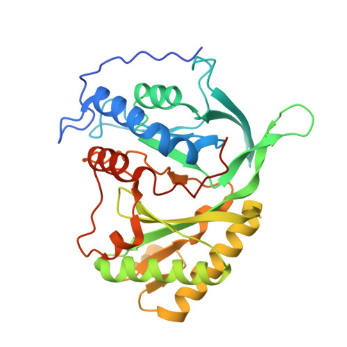
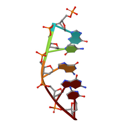
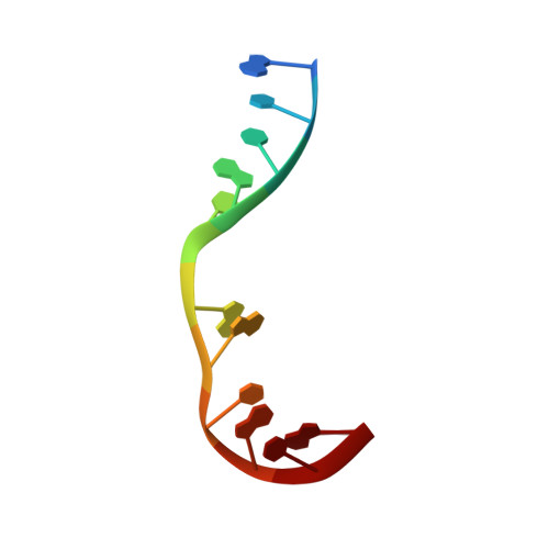
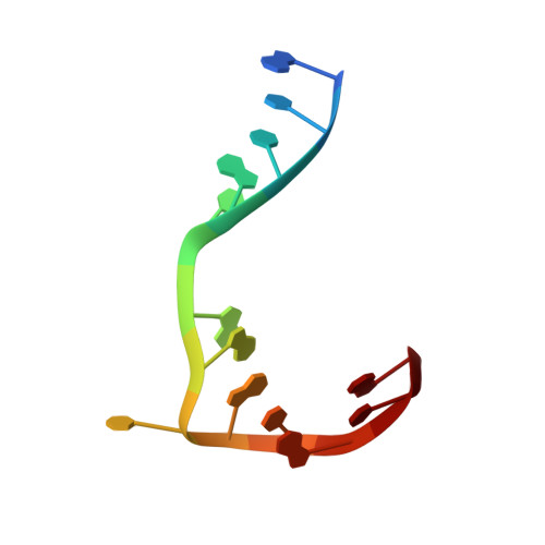
Structure of a NHEJ polymerase-mediated DNA synaptic complex

Brissett, N.C.Pitcher, R.S.Juarez, R.Picher, A.J.Green, A.J.Dafforn, T.R.Fox, G.C.Blanco, L.Doherty, A.J.

(2007) Science 318: 456-459

  • DOI: https://doi.org/10.1126/science.1145112
  • Primary Citation of Related Structures:  
    2R9L

  • PubMed Abstract: 

    Nonhomologous end joining (NHEJ) is a critical DNA double-strand break (DSB) repair pathway required to maintain genome stability. Many prokaryotes possess a minimalist NHEJ apparatus required to repair DSBs during stationary phase, composed of two conserved core proteins, Ku and ligase D (LigD). The crystal structure of Mycobacterium tuberculosis polymerase domain of LigD mediating the synapsis of two noncomplementary DNA ends revealed a variety of interactions, including microhomology base pairing, mismatched and flipped-out bases, and 3' termini forming hairpin-like ends. Biochemical and biophysical studies confirmed that polymerase-induced end synapsis also occurs in solution. We propose that this DNA synaptic structure reflects an intermediate bridging stage of the NHEJ process, before end processing and ligation, with both the polymerase and the DNA sequence playing pivotal roles in determining the sequential order of synapsis and remodeling before end joining.


  • Organizational Affiliation

    Genome Damage and Stability Centre, University of Sussex, Brighton BN1 9RQ, UK.


Macromolecules

Find similar proteins by:  (by identity cutoff)  |  3D Structure
Entity ID: 4
MoleculeChains Sequence LengthOrganismDetailsImage
Putative DNA ligase-like proteinE [auth A],
F [auth B]
303Mycobacterium tuberculosis H37RvMutation(s): 0 
Gene Names: Rv0938
EC: 2.7.7
UniProt
Find proteins for P9WNV3 (Mycobacterium tuberculosis (strain ATCC 25618 / H37Rv))
Explore P9WNV3 
Go to UniProtKB:  P9WNV3
Entity Groups  
Sequence Clusters30% Identity50% Identity70% Identity90% Identity95% Identity100% Identity
UniProt GroupP9WNV3
Sequence Annotations
Expand
  • Reference Sequence

Find similar nucleic acids by:  Sequence   |   3D Structure  

Entity ID: 1
MoleculeChains LengthOrganismImage
DNA (5'-D(P*DGP*DCP*DGP*DGP*DC)-3')A [auth C],
C [auth E]
5synthetic construct
Sequence Annotations
Expand
  • Reference Sequence

Find similar nucleic acids by:  Sequence   |   3D Structure  

Entity ID: 2
MoleculeChains LengthOrganismImage
DNA (5'-D(*DGP*DCP*DCP*DGP*DCP*DAP*DAP*DCP*DGP*DCP*DA)-3')B [auth D]11synthetic construct
Sequence Annotations
Expand
  • Reference Sequence

Find similar nucleic acids by:  Sequence   |   3D Structure  

Entity ID: 3
MoleculeChains LengthOrganismImage
DNA (5'-D(*DGP*DCP*DCP*DGP*DCP*DAP*DAP*DCP*DGP*DCP*DAP*DCP*DG)-3')D [auth F]13synthetic construct
Sequence Annotations
Expand
  • Reference Sequence
Experimental Data & Validation

Experimental Data

  • Method: X-RAY DIFFRACTION
  • Resolution: 2.40 Å
  • R-Value Free: 0.272 
  • R-Value Work: 0.227 
  • R-Value Observed: 0.229 
  • Space Group: P 21 21 21
Unit Cell:
Length ( Å )Angle ( ˚ )
a = 48.531α = 90
b = 111.788β = 90
c = 139.542γ = 90
Software Package:
Software NamePurpose
DENZOdata reduction
SCALEPACKdata scaling
PHASERphasing
REFMACrefinement
PDB_EXTRACTdata extraction

Structure Validation

View Full Validation Report


Entry History 

Deposition Data

Revision History  (Full details and data files)

  • Version 1.0: 2008-01-08
    Type: Initial release
  • Version 1.1: 2011-07-13
    Changes: Advisory, Version format compliance
  • Version 1.2: 2017-10-25
    Changes: Advisory, Refinement description, Source and taxonomy
  • Version 1.3: 2023-08-30
    Changes: Advisory, Data collection, Database references, Derived calculations, Refinement description