2BXF

Human serum albumin complexed with diazepam


Experimental Data Snapshot

  • Method: X-RAY DIFFRACTION
  • Resolution: 2.95 Å
  • R-Value Free: 0.262 
  • R-Value Work: 0.215 
  • R-Value Observed: 0.215 

wwPDB Validation   3D Report Full Report


Ligand Structure Quality Assessment 


This is version 1.3 of the entry. See complete history


Literature

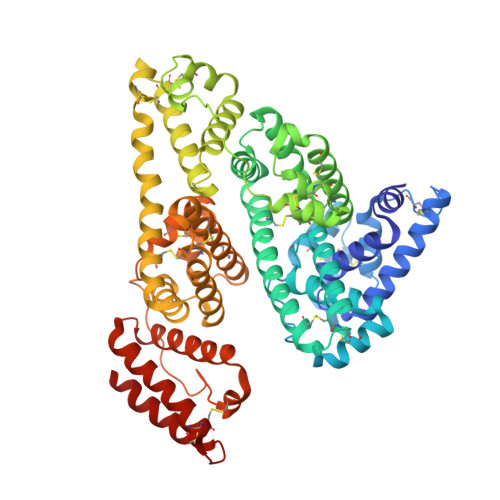
Structural Basis of the Drug-Binding Specificity of Human Serum Albumin.

Ghuman, J.Zunszain, P.A.Petitpas, I.Bhattacharya, A.A.Otagiri, M.Curry, S.

(2005) J Mol Biol 353: 38

  • DOI: https://doi.org/10.1016/j.jmb.2005.07.075
  • Primary Citation of Related Structures:  
    2BX8, 2BXA, 2BXB, 2BXC, 2BXD, 2BXE, 2BXF, 2BXG, 2BXH, 2BXI, 2BXK, 2BXL, 2BXM, 2BXN, 2BXO, 2BXP, 2BXQ

  • PubMed Abstract: 

    Human serum albumin (HSA) is an abundant plasma protein that binds a remarkably wide range of drugs, thereby restricting their free, active concentrations. The problem of overcoming the binding affinity of lead compounds for HSA represents a major challenge in drug development. Crystallographic analysis of 17 different complexes of HSA with a wide variety of drugs and small-molecule toxins reveals the precise architecture of the two primary drug-binding sites on the protein, identifying residues that are key determinants of binding specificity and illuminating the capacity of both pockets for flexible accommodation. Numerous secondary binding sites for drugs distributed across the protein have also been identified. The binding of fatty acids, the primary physiological ligand for the protein, is shown to alter the polarity and increase the volume of drug site 1. These results clarify the interpretation of accumulated drug binding data and provide a valuable template for design efforts to modulate the interaction with HSA.


  • Organizational Affiliation

    Biophysics Section, Division of Cell and Molecular Biology, Imperial College London, South Kensington Campus, London SW7 2AZ, UK.


Macromolecules
Find similar proteins by:  (by identity cutoff)  |  3D Structure
Entity ID: 1
MoleculeChains Sequence LengthOrganismDetailsImage
SERUM ALBUMIN
A, B
585Homo sapiensMutation(s): 0 
UniProt & NIH Common Fund Data Resources
Find proteins for P02768 (Homo sapiens)
Explore P02768 
Go to UniProtKB:  P02768
PHAROS:  P02768
GTEx:  ENSG00000163631 
Entity Groups  
Sequence Clusters30% Identity50% Identity70% Identity90% Identity95% Identity100% Identity
UniProt GroupP02768
Sequence Annotations
Expand
  • Reference Sequence
Small Molecules
Ligands 1 Unique
IDChains Name / Formula / InChI Key2D Diagram3D Interactions
DZP
Query on DZP

Download Ideal Coordinates CCD File 
C [auth A],
D [auth B]
7-CHLORO-1-METHYL-5-PHENYL-1,3-DIHYDRO-2H-1,4-BENZODIAZEPIN-2-ONE
C16 H13 Cl N2 O
AAOVKJBEBIDNHE-UHFFFAOYSA-N
Binding Affinity Annotations 
IDSourceBinding Affinity
DZP BindingDB:  2BXF Kd: 2.45e+4 (nM) from 1 assay(s)
Experimental Data & Validation

Experimental Data

  • Method: X-RAY DIFFRACTION
  • Resolution: 2.95 Å
  • R-Value Free: 0.262 
  • R-Value Work: 0.215 
  • R-Value Observed: 0.215 
  • Space Group: P 1
Unit Cell:
Length ( Å )Angle ( ˚ )
a = 55.29α = 81.17
b = 55.78β = 90.22
c = 120.51γ = 65.93
Software Package:
Software NamePurpose
CNSrefinement
MOSFLMdata reduction
SCALAdata scaling
CNSphasing

Structure Validation

View Full Validation Report


Ligand Structure Quality Assessment 


Entry History 

Deposition Data

Revision History  (Full details and data files)

  • Version 1.0: 2005-09-22
    Type: Initial release
  • Version 1.1: 2011-05-08
    Changes: Version format compliance
  • Version 1.2: 2011-07-13
    Changes: Version format compliance
  • Version 1.3: 2023-12-13
    Changes: Data collection, Database references, Other, Refinement description