1K98

AdoMet complex of MetH C-terminal fragment


Experimental Data Snapshot

  • Method: X-RAY DIFFRACTION
  • Resolution: 3.75 Å
  • R-Value Free: 0.363 
  • R-Value Work: 0.308 
  • R-Value Observed: 0.311 

wwPDB Validation   3D Report Full Report


This is version 1.6 of the entry. See complete history


Literature

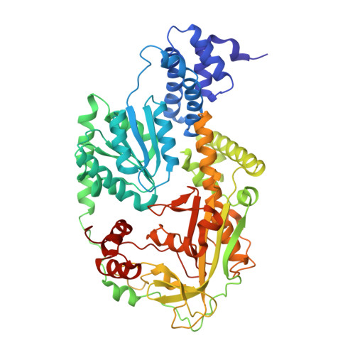
Domain alternation switches B(12)-dependent methionine synthase to the activation conformation.

Bandarian, V.Pattridge, K.A.Lennon, B.W.Huddler, D.P.Matthews, R.G.Ludwig, M.L.

(2002) Nat Struct Biol 9: 53-56

  • DOI: https://doi.org/10.1038/nsb738
  • Primary Citation of Related Structures:  
    1K7Y, 1K98

  • PubMed Abstract: 

    B(12)-dependent methionine synthase (MetH) from Escherichia coli is a large modular protein that uses bound cobalamin as an intermediate methyl carrier. Major domain rearrangements have been postulated to explain how cobalamin reacts with three different substrates: homocysteine, methyltetrahydrofolate and S-adenosylmethionine (AdoMet). Here we describe the 3.0 A structure of a 65 kDa C-terminal fragment of MetH that spans the cobalamin- and AdoMet-binding domains, arranged in a conformation suitable for the methyl transfer from AdoMet to cobalamin that occurs during activation. In the conversion to the activation conformation, a helical domain that capped the cofactor moves 26 A and rotates by 63 degrees, allowing formation of a new interface between cobalamin and the AdoMet-binding (activation) domain. Interactions with the MetH activation domain drive the cobalamin away from its binding domain in a way that requires dissociation of the axial cobalt ligand and, thereby, provide a mechanism for control of the distribution of enzyme conformations.


  • Organizational Affiliation

    Biophysics Research Division and Department of Biological Chemistry, University of Michigan, Ann Arbor, Michigan 48109-1055, USA.


Macromolecules
Find similar proteins by:  (by identity cutoff)  |  3D Structure
Entity ID: 1
MoleculeChains Sequence LengthOrganismDetailsImage
Methionine synthase577Escherichia coliMutation(s): 1 
EC: 2.1.1.13
UniProt
Find proteins for P13009 (Escherichia coli (strain K12))
Explore P13009 
Go to UniProtKB:  P13009
Entity Groups  
Sequence Clusters30% Identity50% Identity70% Identity90% Identity95% Identity100% Identity
UniProt GroupP13009
Sequence Annotations
Expand
  • Reference Sequence
Small Molecules
Ligands 2 Unique
IDChains Name / Formula / InChI Key2D Diagram3D Interactions
B12
Query on B12

Download Ideal Coordinates CCD File 
C [auth A]COBALAMIN
C62 H89 Co N13 O14 P
LKVIQTCSMMVGFU-DWSMJLPVSA-N
SO4
Query on SO4

Download Ideal Coordinates CCD File 
B [auth A]SULFATE ION
O4 S
QAOWNCQODCNURD-UHFFFAOYSA-L
Experimental Data & Validation

Experimental Data

  • Method: X-RAY DIFFRACTION
  • Resolution: 3.75 Å
  • R-Value Free: 0.363 
  • R-Value Work: 0.308 
  • R-Value Observed: 0.311 
  • Space Group: P 43 21 2
Unit Cell:
Length ( Å )Angle ( ˚ )
a = 108.503α = 90
b = 108.503β = 90
c = 146.474γ = 90
Software Package:
Software NamePurpose
CNSrefinement
DENZOdata reduction
SCALEPACKdata scaling
CNSphasing

Structure Validation

View Full Validation Report


Entry History 

Deposition Data

Revision History  (Full details and data files)

  • Version 1.0: 2001-12-21
    Type: Initial release
  • Version 1.1: 2008-04-27
    Changes: Version format compliance
  • Version 1.2: 2011-07-13
    Changes: Version format compliance
  • Version 1.3: 2012-10-24
    Changes: Non-polymer description
  • Version 1.4: 2018-02-14
    Changes: Experimental preparation
  • Version 1.5: 2021-10-27
    Changes: Database references, Derived calculations
  • Version 1.6: 2023-08-16
    Changes: Data collection, Refinement description