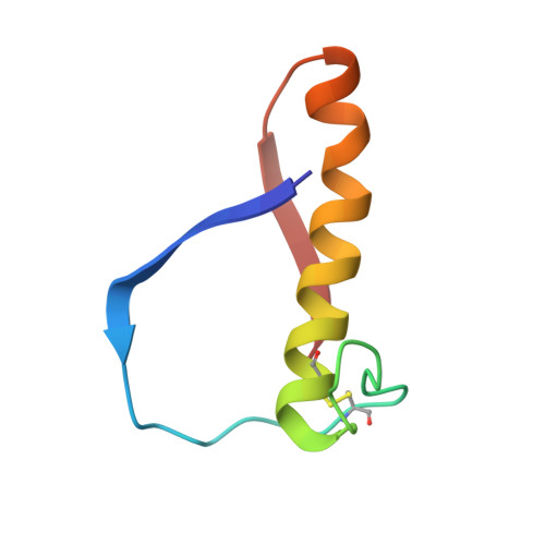
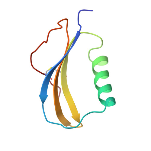
The novel acidophilic structure of the killer toxin from halotolerant yeast demonstrates remarkable folding similarity with a fungal killer toxin.
Kashiwagi, T., Kunishima, N., Suzuki, C., Tsuchiya, F., Nikkuni, S., Arata, Y., Morikawa, K.(1997) Structure 5: 81-94
- PubMed: 9016714
- DOI: https://doi.org/10.1016/s0969-2126(97)00168-8
- Primary Citation of Related Structures:
1KVD, 1KVE - PubMed Abstract:
Several strains of yeasts and fungi produce proteinous substances, termed killer toxins, which kill sensitive strains. The SMK toxin, secreted by the halotolerant yeast Pichia farinosa KK1 strain, uniquely exhibits its maximum killer activity under conditions of acidic pH and high salt concentration. The toxin is composed of two distinct subunits, alpha and beta, which tightly interact with each other under acidic conditions. However, they are easily dissociated under neutral conditions and lose the killer activity. The three-dimensional structure of the SMK toxin will provide a better understanding of the mechanism of toxicity of this protein and the cause of its unique pH-dependent stability. Two crystal structures of the SMK toxin have been determined at 1.8 A resolution in different ionic strength conditions. The two subunits, alpha and beta, are jointly folded into an ellipsoidal, single domain structure belonging to the alpha/beta-sandwich family. The folding topology of the SMK toxin is essentially the same as that of the fungal killer toxin, KP4. This shared topology contains two left-handed split betaalphabeta motifs, which are rare in the other proteins. Many acidic residues are clustered at the bottom of the SMK toxin molecule. Some of the carboxyl sidechains interact with each other through hydrogen bonds. The ionic strength difference induces no evident structural change of the SMK toxin except that, in the high ionic strength crystal, a number of sulfate ions are electrostatically bound near the basic residues which are also locally distributed at the bottom of the toxin molecule. The two killer toxins, SMK and KP4, share a unique folding topology which contains a rare structural motif. This observation may suggest that these toxins are evolutionally and/or functionally related. The pH-dependent stability of the SMK toxin is a result of the intensive interactions between the carboxyl groups. This finding is important for protein engineering, for instance, towards stabilization of the toxin molecule in a broader pH range. The present crystallographic study revealed that the structure of the SMK toxin itself is hardly affected by the ionic strength, implying that a high salt concentration affects the sensitivity of the cell against the toxin.
Organizational Affiliation:
Protein Engineering Research Institute [Biomolecular Engineering Research Institute (BERI) as of the 1st of April 1996], 6-2-3, Furuedai, Suita, Osaka 565, Japan.