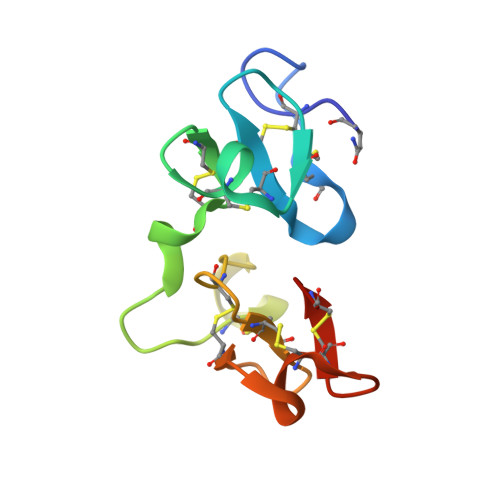
Crystal structure of Urtica dioica agglutinin, a superantigen presented by MHC molecules of class I and class II.
Saul, F.A., Rovira, P., Boulot, G., Damme, E.J., Peumans, W.J., Truffa-Bachi, P., Bentley, G.A.(2000) Structure 8: 593-603
- PubMed: 10873861
- DOI: https://doi.org/10.1016/s0969-2126(00)00142-8
- Primary Citation of Related Structures:
1EIS, 1EN2, 1ENM - PubMed Abstract:
Urtica dioica agglutinin (UDA), a monomeric lectin extracted from stinging nettle rhizomes, is specific for saccharides containing N-acetylglucosamine (GlcNAc). The lectin behaves as a superantigen for murine T cells, inducing the exclusive proliferation of Vbeta8.3(+) lymphocytes. UDA is unique among known T cell superantigens because it can be presented by major histocompatibility complex (MHC) molecules of both class I and II. The crystal structure of UDA has been determined in the ligand-free state, and in complex with tri-acetylchitotriose and tetra-acetylchitotetraose at 1.66 A, 1.90 A and 1.40 A resolution, respectively. UDA comprises two hevein-like domains, each with a saccharide-binding site. A serine and three aromatic residues at each site form the principal contacts with the ligand. The N-terminal domain binding site can centre on any residue of a chito-oligosaccharide, whereas that of the C-terminal domain is specific for residues at the nonreducing terminus of the ligand. We have shown previously that oligomers of GlcNAc inhibit the superantigenic activity of UDA and that the lectin binds to glycans on the MHC molecule. We show that UDA also binds to glycans on the T cell receptor (TCR). The presence of two saccharide-binding sites observed in the structure of UDA suggests that its superantigenic properties arise from the simultaneous fixation of glycans on the TCR and MHC molecules of the T cell and antigen-presenting cell, respectively. The well defined spacing between the two binding sites of UDA is probably a key factor in determining the specificity for Vbeta8.3(+) lymphocytes.
Organizational Affiliation:
Unité d'Immunologie Structurale (CNRS URA 2185), Institut Pasteur, Paris, France.