3JBL | pdb_00003jbl

Cryo-EM Structure of the Activated NAIP2/NLRC4 Inflammasome Reveals Nucleated Polymerization


Experimental Data Snapshot

  • Method: ELECTRON MICROSCOPY
  • Resolution: 4.70 Å
  • Aggregation State: PARTICLE 
  • Reconstruction Method: SINGLE PARTICLE 

wwPDB Validation 3D Report Full Report

Validation slider image for 3JBL

This is version 1.3 of the entry. See complete history

Literature

Cryo-EM structure of the activated NAIP2-NLRC4 inflammasome reveals nucleated polymerization.

Zhang, L.Chen, S.Ruan, J.Wu, J.Tong, A.B.Yin, Q.Li, Y.David, L.Lu, A.Wang, W.L.Marks, C.Ouyang, Q.Zhang, X.Mao, Y.Wu, H.

(2015) Science 350: 404-409

  • DOI: https://doi.org/10.1126/science.aac5789
  • Primary Citation Related Structures: 
    3JBL

  • PubMed Abstract: 

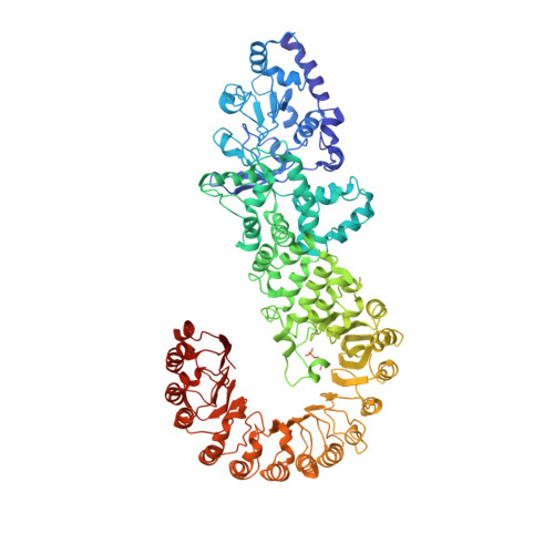
    The NLR family apoptosis inhibitory proteins (NAIPs) bind conserved bacterial ligands, such as the bacterial rod protein PrgJ, and recruit NLR family CARD-containing protein 4 (NLRC4) as the inflammasome adapter to activate innate immunity. We found that the PrgJ-NAIP2-NLRC4 inflammasome is assembled into multisubunit disk-like structures through a unidirectional adenosine triphosphatase polymerization, primed with a single PrgJ-activated NAIP2 per disk. Cryo-electron microscopy (cryo-EM) reconstruction at subnanometer resolution revealed a ~90° hinge rotation accompanying NLRC4 activation. Unlike in the related heptameric Apaf-1 apoptosome, in which each subunit needs to be conformationally activated by its ligand before assembly, a single PrgJ-activated NAIP2 initiates NLRC4 polymerization in a domino-like reaction to promote the disk assembly. These insights reveal the mechanism of signal amplification in NAIP-NLRC4 inflammasomes.


  • Organizational Affiliation
    • Department of Biological Chemistry and Molecular Pharmacology, Harvard Medical School, Boston, MA 02115, USA. Program in Cellular and Molecular Medicine, Boston Children's Hospital, Boston, MA 02115, USA.

Macromolecule Content 

  • Total Structure Weight: 1,168.64 kDa 
  • Atom Count: 80,124 
  • Modeled Residue Count: 9,999 
  • Deposited Residue Count: 10,252 
  • Unique protein chains: 1

Macromolecules

Find similar proteins by:|  3D Structure
Entity ID: 1
MoleculeChains  Sequence LengthOrganismDetailsImage
NLR family CARD domain-containing protein 4932Mus musculusMutation(s): 0 
Gene Names: Nlrc4Card12Ipaf
UniProt
Find proteins for Q3UP24 (Mus musculus)
Explore Q3UP24 
Go to UniProtKB:  Q3UP24
Entity Groups
Sequence Clusters30% Identity50% Identity70% Identity90% Identity95% Identity100% Identity
UniProt GroupQ3UP24
Sequence Annotations
Expand
Reference Sequence

Small Molecules

Modified Residues  1 Unique
IDChains TypeFormula2D DiagramParent
SEP
Query on SEP
A [auth K]
B [auth A]
C [auth B]
D [auth C]
E [auth D]
A [auth K],
B [auth A],
C [auth B],
D [auth C],
E [auth D],
F [auth E],
G [auth F],
H [auth G],
I [auth H],
J [auth I],
K [auth J]
L-PEPTIDE LINKINGC3 H8 N O6 PSER

Experimental Data & Validation

Experimental Data

  • Method: ELECTRON MICROSCOPY
  • Resolution: 4.70 Å
  • Aggregation State: PARTICLE 
  • Reconstruction Method: SINGLE PARTICLE 
EM Software:
TaskSoftware PackageVersion
RECONSTRUCTIONEMAN2
RECONSTRUCTIONRELION
RECONSTRUCTIONSPIDER

Structure Validation

View Full Validation Report


Entry History 

Deposition Data

Revision History  (Full details and data files)

  • Version 1.0: 2015-10-21
    Type: Initial release
  • Version 1.1: 2015-11-04
    Changes: Database references, Other
  • Version 1.2: 2018-07-18
    Changes: Data collection
  • Version 1.3: 2024-11-20
    Changes: Data collection, Database references, Derived calculations, Refinement description, Structure summary