


Crystal Structure of 6,7-dimethyl-8-ribityllumazine synthase from Bordetella pertussis in complex with 6,7-dimethyl-8-(1'-D-ribityl) lumazine
Seibold, S., Lovell, S., Battaile, K.P.To be published.
Experimental Data Snapshot
Starting Model: experimental
View more details
Entity ID: 1 | |||||
|---|---|---|---|---|---|
| Molecule | Chains | Sequence Length | Organism | Details | Image |
| 6,7-dimethyl-8-ribityllumazine synthase | 163 | Bordetella pertussis Tohama I | Mutation(s): 0 Gene Names: ribH, BP3485 EC: 2.5.1.78 | 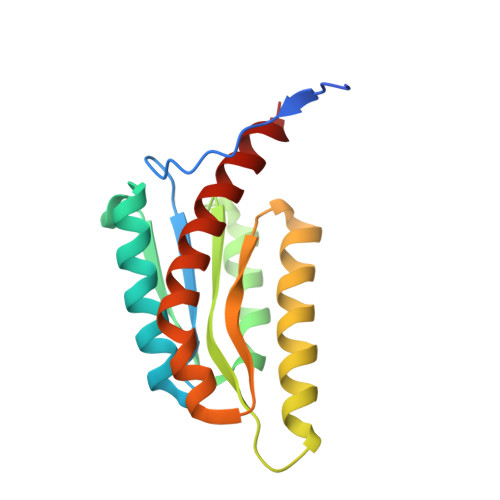 | |
UniProt | |||||
Entity Groups | |||||
| Sequence Clusters | 30% Identity50% Identity70% Identity90% Identity95% Identity100% Identity | ||||
| UniProt Group | Q7VTN4 | ||||
Sequence AnnotationsExpand | |||||
Reference Sequence | |||||
| Ligands 3 Unique | |||||
|---|---|---|---|---|---|
| ID | Chains | Name / Formula / InChI Key | 2D Diagram | 3D Interactions | |
| DLZ (Subject of Investigation/LOI) Download:Ideal Coordinates CCD File | AA [auth D], FA [auth E], L [auth A], Q [auth B], W [auth C] | 1-deoxy-1-(6,7-dimethyl-2,4-dioxo-3,4-dihydropteridin-8(2H)-yl)-D-ribitol C13 H18 N4 O6 SXDXRJZUAJBNFL-XKSSXDPKSA-N |  | ||
| PO4 Download:Ideal Coordinates CCD File | BA [auth E] CA [auth E] F [auth A] M [auth B] R [auth C] | PHOSPHATE ION O4 P NBIIXXVUZAFLBC-UHFFFAOYSA-K |  | ||
| CL Download:Ideal Coordinates CCD File | DA [auth E] EA [auth E] G [auth A] H [auth A] I [auth A] | CHLORIDE ION Cl VEXZGXHMUGYJMC-UHFFFAOYSA-M |  | ||
| Length ( Å ) | Angle ( ˚ ) |
|---|---|
| a = 135.448 | α = 90 |
| b = 75.416 | β = 114.38 |
| c = 94.461 | γ = 90 |
| Software Name | Purpose |
|---|---|
| PHENIX | refinement |
| Aimless | data scaling |
| XDS | data reduction |
| PHASER | phasing |
| PDB_EXTRACT | data extraction |
| Funding Organization | Location | Grant Number |
|---|---|---|
| National Institutes of Health/National Institute Of Allergy and Infectious Diseases (NIH/NIAID) | United States | 75N93022C00036 |
| National Institutes of Health/Office of the Director | United States | S10OD030394 |


