A Novel Small Molecule Allosteric Inhibitor of IL-17A from a DNA-Encoded Library.
Milla, M.E., Blevitt, J.M., Goldberg, S.D., Armstrong, A.A., Blain, K.Y., Herman, K.L., Liu, A.X., Luna, R., Milligan, C.M., Patrick, A.N., Steele, R.A., Bembenek, S.D., Centrella, P., Clark, M.A., Cuozzo, J.W., Disch, J.S., Domingo, D., Hunt, A., Hupp, C.D., Keefe, A.D., Luo, J., Mirzadegan, T., Nelen, M.I., Resnicow, D.I., Sigel, E.A., Soutter, H.H., Troast, D.M., Xue, X., Yi, F., Zhang, Y., Jackson, P.F., Edwards, J.P., Lumb, K.J.(2025) ACS Med Chem Lett 16: 2301-2308
- PubMed: 41256970 Search on PubMedSearch on PubMed Central
- DOI: https://doi.org/10.1021/acsmedchemlett.5c00502
- Primary Citation Related Structures:
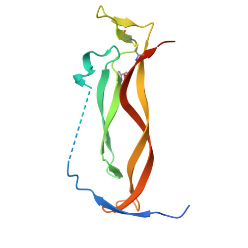
9YIC - PubMed Abstract:
A novel series of inhibitors of the interaction of IL-17A with its cognate receptor has been discovered using DNA-encoded library (DEL) technology. The lead compound (JNJ627, Compound 1 ) of the series occupies the interior interface of the IL-17A homodimer and disables receptor binding. The mechanism of action involves allosteric disruption of the IL-17A quaternary structure to prevent adoption of the receptor-binding conformation, rather than direct orthosteric inhibition at the receptor-binding site. Molecules of this series exhibit remarkably slow on-rate kinetics and potent inhibition of IL-17A signaling in human primary cells.
- Johnson & Johnson Innovative Medicine, 3210 Merryfield Row, La Jolla, California 92121, United States.
Organizational Affiliation: