9YDU | pdb_00009ydu

UDPG bound P2Y14 Receptor in complex with Gi


Experimental Data Snapshot

  • Method: ELECTRON MICROSCOPY
  • Resolution: 3.19 Å
  • Aggregation State: PARTICLE 
  • Reconstruction Method: SINGLE PARTICLE 

wwPDB Validation   3D Report Full Report


This is version 1.2 of the entry. See complete history


Literature

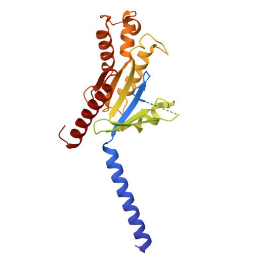
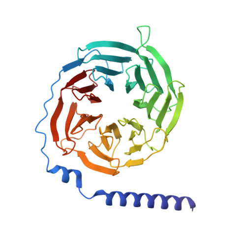
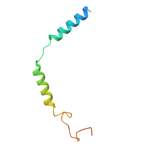
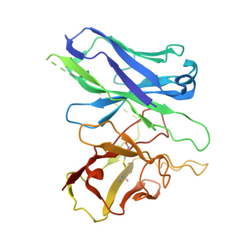
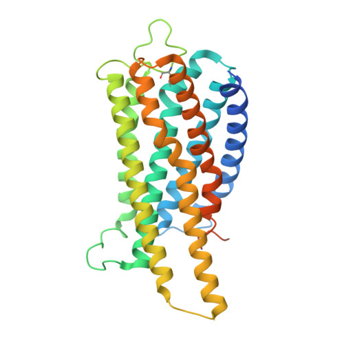
UDP-glucose and MRS2905 agonist-bound states of the purinergic P2Y 14 receptor.

Fay, J.F.Kousouros, J.Gao, Z.G.Pavan, M.Oliva, P.Pramanik, A.Meyer, C.Kurma, S.H.Xu, W.Krumm, B.Jacobson, K.A.

(2025) Commun Biol 8: 1816-1816

  • DOI: https://doi.org/10.1038/s42003-025-09174-6
  • Primary Citation of Related Structures:  
    9YDU, 9YDV

  • PubMed Abstract: 

    P2Y 14 receptors respond to uracil-diphosphate-hexose conjugates, yet how this receptor selectively recognizes both uracil-nucleotides and hexose moieties of diverse agonists remains unclear. Here we report the active agonist-bound G protein-bound states of the P2Y 14 G protein-coupled receptor (GPCR) bound to endogenous agonist uridine diphosphate glucose (UDP-glucose) and 2-thiouridine-5'-O-(α,β-methylene)diphosphate (MRS2905). The cryo-EM structures of the heterotrimeric complexes with 3.19 Å and 3.05 Å global resolution, respectively, with local refinements reaching 2.87 Å and 3.22 Å for the masked receptor region. Our structures reveal a pronounced extracellular facing electronegative vestibule connecting to a smaller nucleotide binding subpocket (~300Å 3 volume) that is shielded by extracellular loop 2 (ECL2). A glucose-binding subpocket is spatially delimited by residue V93; mutation to Trp selectively blocks UDP-glucose while permitting MRS2905 and antagonist binding. These findings provide atomic insights into uracil recognition, reveal how the receptor accommodates diverse flexible UDP-sugars, and promise to enable rational drug discovery of therapeutic P2Y 14 R modulators.


  • Organizational Affiliation
    • Department of Biochemistry and Molecular Biology, University of Maryland Baltimore, Baltimore, MD, USA. jfay@som.umaryland.edu.

Macromolecules
Find similar proteins by:  (by identity cutoff)  |  3D Structure
Entity ID: 1
MoleculeChains Sequence LengthOrganismDetailsImage
Guanine nucleotide-binding protein G(i) subunit alpha-1A [auth B]354Homo sapiensMutation(s): 4 
Gene Names: GNAI1
EC: 3.6.5
UniProt & NIH Common Fund Data Resources
Find proteins for P63096 (Homo sapiens)
Explore P63096 
Go to UniProtKB:  P63096
PHAROS:  P63096
GTEx:  ENSG00000127955 
Entity Groups  
Sequence Clusters30% Identity50% Identity70% Identity90% Identity95% Identity100% Identity
UniProt GroupP63096
Sequence Annotations
Expand
  • Reference Sequence
Find similar proteins by:  (by identity cutoff)  |  3D Structure
Entity ID: 2
MoleculeChains Sequence LengthOrganismDetailsImage
Guanine nucleotide-binding protein G(I)/G(S)/G(T) subunit beta-1B [auth C]340Homo sapiensMutation(s): 0 
Gene Names: GNB1
UniProt & NIH Common Fund Data Resources
Find proteins for P62873 (Homo sapiens)
Explore P62873 
Go to UniProtKB:  P62873
PHAROS:  P62873
GTEx:  ENSG00000078369 
Entity Groups  
Sequence Clusters30% Identity50% Identity70% Identity90% Identity95% Identity100% Identity
UniProt GroupP62873
Sequence Annotations
Expand
  • Reference Sequence
Find similar proteins by:  (by identity cutoff)  |  3D Structure
Entity ID: 3
MoleculeChains Sequence LengthOrganismDetailsImage
Guanine nucleotide-binding protein G(I)/G(S)/G(O) subunit gamma-2C [auth D]71Homo sapiensMutation(s): 0 
Gene Names: GNG2
UniProt & NIH Common Fund Data Resources
Find proteins for P59768 (Homo sapiens)
Explore P59768 
Go to UniProtKB:  P59768
PHAROS:  P59768
GTEx:  ENSG00000186469 
Entity Groups  
Sequence Clusters30% Identity50% Identity70% Identity90% Identity95% Identity100% Identity
UniProt GroupP59768
Sequence Annotations
Expand
  • Reference Sequence
Find similar proteins by:  (by identity cutoff)  |  3D Structure
Entity ID: 4
MoleculeChains Sequence LengthOrganismDetailsImage
scFv16D [auth E]251Mus musculusMutation(s): 0 
Entity Groups  
Sequence Clusters30% Identity50% Identity70% Identity90% Identity95% Identity100% Identity
Sequence Annotations
Expand
  • Reference Sequence
Find similar proteins by:  (by identity cutoff)  |  3D Structure
Entity ID: 5
MoleculeChains Sequence LengthOrganismDetailsImage
P2Y purinoceptor 14E [auth A]338Homo sapiensMutation(s): 0 
Gene Names: P2RY14GPR105KIAA0001
UniProt & NIH Common Fund Data Resources
Find proteins for Q15391 (Homo sapiens)
Explore Q15391 
Go to UniProtKB:  Q15391
PHAROS:  Q15391
GTEx:  ENSG00000174944 
Entity Groups  
Sequence Clusters30% Identity50% Identity70% Identity90% Identity95% Identity100% Identity
UniProt GroupQ15391
Sequence Annotations
Expand
  • Reference Sequence
Small Molecules
Ligands 1 Unique
IDChains Name / Formula / InChI Key2D Diagram3D Interactions
UPG (Subject of Investigation/LOI)
Query on UPG

Download Ideal Coordinates CCD File 
F [auth A]URIDINE-5'-DIPHOSPHATE-GLUCOSE
C15 H24 N2 O17 P2
HSCJRCZFDFQWRP-JZMIEXBBSA-N
Experimental Data & Validation

Experimental Data

  • Method: ELECTRON MICROSCOPY
  • Resolution: 3.19 Å
  • Aggregation State: PARTICLE 
  • Reconstruction Method: SINGLE PARTICLE 
EM Software:
TaskSoftware PackageVersion
RECONSTRUCTIONcryoSPARC3.2

Structure Validation

View Full Validation Report


Entry History & Funding Information

Deposition Data

  • Released Date: 2025-11-19 
  • Deposition Author(s): Fay, J.F.

Funding OrganizationLocationGrant Number
National Institutes of Health/National Institute of General Medical Sciences (NIH/NIGMS)United States--

Revision History  (Full details and data files)

  • Version 1.0: 2025-11-19
    Type: Initial release
  • Version 1.1: 2025-12-03
    Changes: Data collection, Database references
  • Version 1.2: 2026-01-07
    Changes: Data collection, Database references