9Y9W | pdb_00009y9w

Vibrio cholerae protein FrhA peptid-binding domain and adjacent split domain (S1127-F1439) in complex with peptide AGWTD X-ray crystallography structure


Experimental Data Snapshot

  • Method: X-RAY DIFFRACTION
  • Resolution: 2.40 Å
  • R-Value Free: 
    0.232 (Depositor), 0.232 (DCC) 
  • R-Value Work: 
    0.227 (Depositor), 0.229 (DCC) 
  • R-Value Observed: 
    0.227 (Depositor) 

Starting Model: experimental
View more details

wwPDB Validation   3D Report Full Report


This is version 1.0 of the entry. See complete history


Literature

Peptide-based ligand antagonists block a Vibrio cholerae adhesin.

Wang, M.Du, G.Yongo-Luwawa, C.Lu, A.Kinrade, B.Munro, K.Klose, K.E.Lubell, W.D.Davies, P.Guo, S.

(2025) FEBS Lett 

  • DOI: https://doi.org/10.1002/1873-3468.70231
  • Primary Citation of Related Structures:  
    9Y9W, 9YBQ

  • PubMed Abstract: 

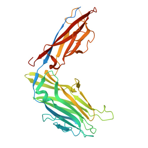
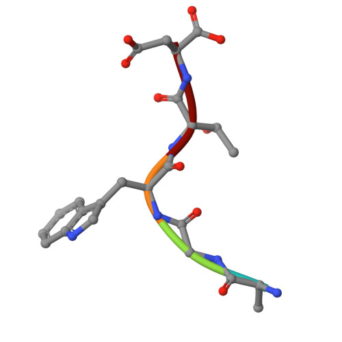
    Vibrio cholerae, the causative agent of cholera, uses surface proteins such as the repeats-in-toxin (RTX) adhesin FrhA to colonize hosts and initiate infection. Blocking bacterial adhesion represents a promising therapeutic strategy to treat infections without promoting drug resistance. FrhA contains a peptide-binding domain (PBD) that is key for hemagglutination, human epithelial cell binding, and V. cholerae biofilm formation. Previous studies identified a lead pentapeptide ligand with the sequence Ala-Gly-Tyr-Thr-Asp (AGYTD) that blocks V. cholerae colonization of the mouse small intestine at high micromolar concentrations. In this study, a structure-guided approach identified a minimal D-amino acid-containing tripeptide motif with higher affinity for the FrhA-PBD and predicted metabolic stability. Our results contribute to the development of anti-adhesion strategies to combat infections. Impact statement Our study elucidates the molecular basis of peptide recognition by the Vibrio cholerae adhesin FrhA and develops minimal D-amino-acid peptides that block adhesion with nanomolar affinity. These findings advance understanding of RTX adhesins and provide a structural blueprint for next-generation anti-adhesion therapeutics against cholera and related infections.


  • Organizational Affiliation
    • Department of Anatomy and Cell Biology, McGill University, Montreal, Canada.

Macromolecules
Find similar proteins by:  (by identity cutoff)  |  3D Structure
Entity ID: 1
MoleculeChains Sequence LengthOrganismDetailsImage
Cadherin domain protein363Vibrio cholerae O395Mutation(s): 0 
Gene Names: VC0395_A1227
UniProt
Find proteins for A0A0H3AMP4 (Vibrio cholerae serotype O1 (strain ATCC 39541 / Classical Ogawa 395 / O395))
Explore A0A0H3AMP4 
Go to UniProtKB:  A0A0H3AMP4
Entity Groups  
Sequence Clusters30% Identity50% Identity70% Identity90% Identity95% Identity100% Identity
UniProt GroupA0A0H3AMP4
Sequence Annotations
Expand
  • Reference Sequence

Find similar proteins by:  Sequence   |   3D Structure  

Entity ID: 2
MoleculeChains Sequence LengthOrganismDetailsImage
Ala-Gly-Trp-Thr-Asp (AGWTD)5Vibrio choleraeMutation(s): 0 
Sequence Annotations
Expand
  • Reference Sequence
Small Molecules
Ligands 3 Unique
IDChains Name / Formula / InChI Key2D Diagram3D Interactions
PEG
Query on PEG

Download Ideal Coordinates CCD File 
PA [auth G],
Y [auth B]
DI(HYDROXYETHYL)ETHER
C4 H10 O3
MTHSVFCYNBDYFN-UHFFFAOYSA-N
EDO
Query on EDO

Download Ideal Coordinates CCD File 
AA [auth B],
BA [auth B],
IA [auth E],
WA [auth I],
Z [auth B]
1,2-ETHANEDIOL
C2 H6 O2
LYCAIKOWRPUZTN-UHFFFAOYSA-N
CA (Subject of Investigation/LOI)
Query on CA

Download Ideal Coordinates CCD File 
AB [auth K]
BB [auth K]
CA [auth E]
CB [auth K]
DA [auth E]
AB [auth K],
BB [auth K],
CA [auth E],
CB [auth K],
DA [auth E],
EA [auth E],
FA [auth E],
GA [auth E],
HA [auth E],
JA [auth G],
KA [auth G],
LA [auth G],
M [auth A],
MA [auth G],
N [auth A],
NA [auth G],
O [auth A],
OA [auth G],
P [auth A],
Q [auth A],
QA [auth I],
R [auth A],
RA [auth I],
S [auth B],
SA [auth I],
T [auth B],
TA [auth I],
U [auth B],
UA [auth I],
V [auth B],
VA [auth I],
W [auth B],
X [auth B],
XA [auth K],
YA [auth K],
ZA [auth K]
CALCIUM ION
Ca
BHPQYMZQTOCNFJ-UHFFFAOYSA-N
Experimental Data & Validation

Experimental Data

  • Method: X-RAY DIFFRACTION
  • Resolution: 2.40 Å
  • R-Value Free:  0.232 (Depositor), 0.232 (DCC) 
  • R-Value Work:  0.227 (Depositor), 0.229 (DCC) 
  • R-Value Observed: 0.227 (Depositor) 
Space Group: P 1
Unit Cell:
Length ( Å )Angle ( ˚ )
a = 61.273α = 60.03
b = 118.726β = 85.62
c = 118.706γ = 76.7
Software Package:
Software NamePurpose
PHENIXrefinement
xia2data reduction
DIALSdata scaling
PHENIXphasing
Cootmodel building

Structure Validation

View Full Validation Report


Entry History & Funding Information

Deposition Data


Funding OrganizationLocationGrant Number
Fonds de Recherche du Quebec - Sante (FRQS)Canada359456
Fonds de Recherche du Quebec - Sante (FRQS)Canada376506
Natural Sciences and Engineering Research Council (NSERC, Canada)CanadaRGPIN-2024-04631
Canadian Institutes of Health Research (CIHR)CanadaFRN 148422

Revision History  (Full details and data files)

  • Version 1.0: 2025-12-03
    Type: Initial release