9X2O | pdb_00009x2o

X-ray structure of antiviral protein from Mirabilis jalapa


Experimental Data Snapshot

  • Method: X-RAY DIFFRACTION
  • Resolution: 2.10 Å
  • R-Value Free: 
    0.246 (Depositor), 0.247 (DCC) 
  • R-Value Work: 
    0.190 (Depositor), 0.191 (DCC) 
  • R-Value Observed: 
    0.193 (Depositor) 

Starting Model: in silico
View more details

wwPDB Validation 3D Report Full Report

Validation slider image for 9X2O

Ligand Structure Quality Assessment 


This is version 1.2 of the entry. See complete history

Literature

Structure of Ribosome-Inactivating Protein from Mirabilis jalapa and Its L12-Stalk-Dependent Inhibition of Escherichia coli Ribosome.

Nishida, N.Ninomiya, Y.Yoshida, T.Tanzawa, T.Maki, Y.Yoshida, H.Tsuge, H.Habuka, N.

(2025) Toxins (Basel) 17

  • DOI: https://doi.org/10.3390/toxins17120575
  • Primary Citation Related Structures: 
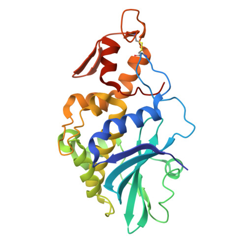
    9X2O

  • PubMed Abstract: 

    Mirabilis antiviral protein (MAP) is the type I ribosome-inactivating protein (RIP), which consists of an RNA N -glycosylase domain with no carbohydrate-binding domain. Unlike many RIPs, such as ricin or trichosanthin, which inactivate eukaryotic ribosomes, MAP also inactivates the E. coli ribosome by cleaving the N -glycosidic bond at A2660 of 23S ribosomal RNA. The structure of the wild-type MAP has not been revealed yet. Here, we expressed, purified, and crystallized the plural recombinant MAPs, including both E168Q and R171Q mutations (MAP-EQRQ) in E. coli , and determined the crystal structure of MAP-EQRQ at 2.1 Å resolution. According to the predicted structure with RNA (sarcin-ricin loop) and the mutant protein's activities using quantitative RT-PCR, we showed that residue R171 at the active site of MAP is a key residue to form the stable complex with target adenine. Furthermore, we showed that MAP bound the C-terminal domains of eukaryotic P2-stalk as well as E. coli L12-stalk.


  • Organizational Affiliation
    • Faculty of Life Sciences, Kyoto Sangyo University, Kita-ku, Kyoto 603-8555, Japan.

Macromolecule Content 

  • Total Structure Weight: 115.9 kDa 
  • Atom Count: 8,723 
  • Modeled Residue Count: 1,004 
  • Deposited Residue Count: 1,024 
  • Unique protein chains: 1

Macromolecules

Find similar proteins by:|  3D Structure
Entity ID: 1
MoleculeChains  Sequence LengthOrganismDetailsImage
Antiviral protein MAP
A, B, C, D
256Mirabilis jalapaMutation(s): 0 
EC: 3.2.2.22
UniProt
Find proteins for P21326 (Mirabilis jalapa)
Explore P21326 
Go to UniProtKB:  P21326
Entity Groups
Sequence Clusters30% Identity50% Identity70% Identity90% Identity95% Identity100% Identity
UniProt GroupP21326
Sequence Annotations
Expand
Reference Sequence

Small Molecules

Ligands 1 Unique
IDChains Name / Formula / InChI Key2D Diagram3D Interactions
SO4
(Subject of Investigation/LOI)

Query on SO4


Download:Ideal Coordinates CCD File
E [auth A]
F [auth B]
G [auth B]
H [auth B]
I [auth C]
E [auth A],
F [auth B],
G [auth B],
H [auth B],
I [auth C],
J [auth C],
K [auth C],
L [auth C],
M [auth C],
N [auth D],
O [auth D],
P [auth D],
Q [auth D]
SULFATE ION
O4 S
QAOWNCQODCNURD-UHFFFAOYSA-L

Experimental Data & Validation

Experimental Data

  • Method: X-RAY DIFFRACTION
  • Resolution: 2.10 Å
  • R-Value Free:  0.246 (Depositor), 0.247 (DCC) 
  • R-Value Work:  0.190 (Depositor), 0.191 (DCC) 
  • R-Value Observed: 0.193 (Depositor) 
Space Group: C 1 2 1
Unit Cell:
Length ( Å )Angle ( ˚ )
a = 214.312α = 90
b = 60.645β = 109.02
c = 79.254γ = 90
Software Package:
Software NamePurpose
PHENIXrefinement
XDSdata reduction
XDSdata scaling
PHENIXphasing

Structure Validation

View Full Validation Report


Ligand Structure Quality Assessment 


Entry History 

& Funding Information

Deposition Data


Funding OrganizationLocationGrant Number
Japan Society for the Promotion of Science (JSPS)Japan23K18011

Revision History  (Full details and data files)

  • Version 1.0: 2025-12-03
    Type: Initial release
  • Version 1.1: 2025-12-10
    Changes: Database references
  • Version 1.2: 2026-01-07
    Changes: Database references