Long lasting complement neutralisation by RAY121, an engineered anti-C1s antibody with C1q displacement function.
Ho, A.W.S., Fukuzawa, T., Koga, H., Ohmine, K., Kawauchi, H., Muraoka, M., Ikuta, Y., Gupta, G., Okuda, M., Onami, I., Ayabe, M., Takeiri, A., Konishi, H., Sugawara, Y., Usami, S., Ito, E., Shibahara, N., Ozeki, K., Wada, Y., Hirako, A., Takahashi, N., Sampei, Z., Haraya, K., Murao, N., Fujii, T., Torizawa, T., Shimada, H., Igawa, T.(2025) EBioMedicine 122: 106009-106009
- PubMed: 41205294 Search on PubMed
- DOI: https://doi.org/10.1016/j.ebiom.2025.106009
- Primary Citation Related Structures:
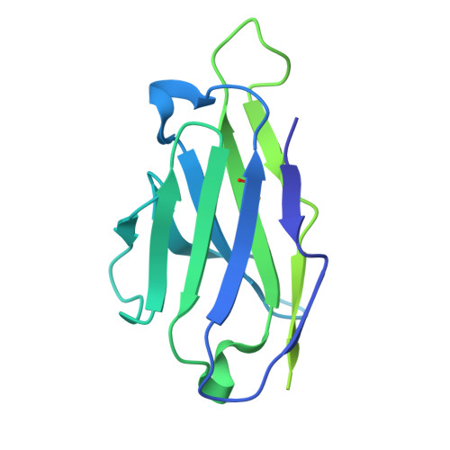
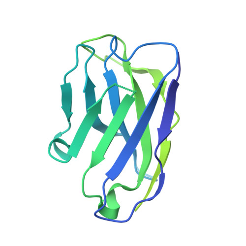
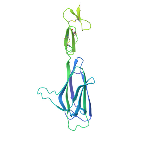
9X1H - PubMed Abstract:
Antibody drugs blocking the complement classical pathway (CP) often require frequent intravenous administration to overcome the high abundance or rapid turnover of complement proteins. This makes treatment compliance burdensome for patients. Screening was performed to identify anti-C1s antibodies specific for the CUB domain of C1s with CP neutralising function. Antibody engineering was performed on the anti-C1s antibody to prolong its half-life in circulation. The variable region was mutated to confer pH-dependent binding to C1s, and the antibody Fc was modified to lower its isoelectric point and to have enhanced binding to the neonatal Fc receptor. A combination of pH-dependent binding to C1s and optimisation of charge characteristics was required for the final engineered antibody, RAY121, to achieve a long half-life in circulation and long-lasting neutralisation of the CP. In male cynomolgus monkeys, a single subcutaneous injection suppressed CP activity for more than a month. RAY121 neutralises the CP by using a unique mechanism of displacing C1q from the C1 complex and blocking its reassembly. Structure analysis by cryo-electron microscopy revealed that the RAY121 epitope on C1s is distinct from the C1q binding site, and that C1q displacement is mediated by the Fab arm sterically obstructing C1q binding to C1s. RAY121 is able to neutralise the CP for an extended duration and is effective at doses that can be administered subcutaneously for convenient treatment. These data present RAY121 as a promising agent for the treatment of CP-driven autoimmune disorders. Chugai Pharmaceutical Co. Ltd.
- Chugai Pharmabody Research, Singapore, 138623, Singapore. Electronic address: adrian.h@chugai-pharmabody.com.
Organizational Affiliation: