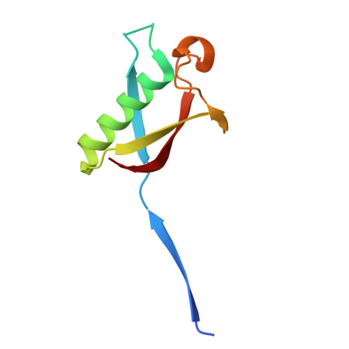
9WVH | pdb_00009wvh

Crystal structure of TONSL UBL domain


Experimental Data Snapshot

  • Method: X-RAY DIFFRACTION
  • Resolution: 1.96 Å
  • R-Value Free: 
    0.209 (Depositor), 0.219 (DCC) 
  • R-Value Work: 
    0.194 (Depositor), 0.204 (DCC) 
  • R-Value Observed: 
    0.195 (Depositor) 

Starting Model: in silico
View more details

wwPDB Validation 3D Report Full Report

Validation slider image for 9WVH

This is version 1.1 of the entry. See complete history

Literature

Pathogenic variants in the human TONSL protein associated with SPONASTRIME dysplasia impair protein dimerization and DNA repair.

Karmakar, A.Roy, S.

(2026) Sci Adv 12: eaee1129-eaee1129

  • DOI: https://doi.org/10.1126/sciadv.aee1129
  • Primary Citation Related Structures: 
    9WVH, 9WVI

  • PubMed Abstract: 

    TONSL safeguards genome stability by facilitating replication-dependent DNA damage repair and protecting stalled replication forks through homologous recombination. Mutations in TONSL cause SPONASTRIME dysplasia, a rare skeletal disorder. We reveal that TONSL homo-dimerizes via its ubiquitin-like domain (UBL), and two recurrent SPONASTRIME dysplasia causative variants (R934W and G973R) abolish this dimerization. Crystal structures at 1.9 Å resolution show UBL WT forms domain-swapped dimers that assemble into ring-like octamers. The R934W variant eliminates critical hydrogen bonds and introduces steric clashes, forcing monomeric conformation. G973R destabilizes an evolutionarily conserved residue within a conformationally restricted β-turn. Biochemically, UBL WT exists as dimers while UBL R934W remains monomeric. Functionally, dimerization-deficient variants fail to suppress replication stress-induced DNA damage, show impaired RAD51 foci formation, and exhibit severely compromised survival following genotoxic stress. These findings establish TONSL dimerization as essential for genome maintenance and provide structural and mechanistic insights into SPONASTRIME dysplasia pathogenesis.


  • Organizational Affiliation
    • Structural Biology and Bio-Informatics Division, Council of Scientific & Industrial Research-Indian Institute of Chemical Biology, Kolkata-700032, India.

Macromolecule Content 

  • Total Structure Weight: 19.56 kDa 
  • Atom Count: 1,378 
  • Modeled Residue Count: 159 
  • Deposited Residue Count: 170 
  • Unique protein chains: 1

Macromolecules

Find similar proteins by:|  3D Structure
Entity ID: 1
MoleculeChains  Sequence LengthOrganismDetailsImage
Tonsoku-like protein
A, B
85Homo sapiensMutation(s): 0 
Gene Names: TONSLIKBRNFKBIL2
UniProt & NIH Common Fund Data Resources
Find proteins for Q96HA7 (Homo sapiens)
Explore Q96HA7 
Go to UniProtKB:  Q96HA7
GTEx:  ENSG00000160949 
Entity Groups
Sequence Clusters30% Identity50% Identity70% Identity90% Identity95% Identity100% Identity
UniProt GroupQ96HA7
Sequence Annotations
Expand
Reference Sequence

Small Molecules

Ligands 1 Unique
IDChains Name / Formula / InChI Key2D Diagram3D Interactions
NA

Query on NA


Download:Ideal Coordinates CCD File
C [auth A]SODIUM ION
Na
FKNQFGJONOIPTF-UHFFFAOYSA-N

Experimental Data & Validation

Experimental Data

  • Method: X-RAY DIFFRACTION
  • Resolution: 1.96 Å
  • R-Value Free:  0.209 (Depositor), 0.219 (DCC) 
  • R-Value Work:  0.194 (Depositor), 0.204 (DCC) 
  • R-Value Observed: 0.195 (Depositor) 
Space Group: I 4 2 2
Unit Cell:
Length ( Å )Angle ( ˚ )
a = 112.147α = 90
b = 112.147β = 90
c = 69.278γ = 90
Software Package:
Software NamePurpose
REFMACrefinement
PROTEUM PLUSdata collection
XDSdata reduction
XSCALEdata scaling
PHASERphasing

Structure Validation

View Full Validation Report


Entry History 

& Funding Information

Deposition Data


Funding OrganizationLocationGrant Number
Science and Engineering Research Board (SERB)IndiaCRG/2022/001895

Revision History  (Full details and data files)

  • Version 1.0: 2025-10-08
    Type: Initial release
  • Version 1.1: 2026-08-12
    Changes: Database references