


Crystal structure of human ZMYND8 MYND domain
Srivastava, D.K., Roy, S.To be published.
Experimental Data Snapshot
Starting Model: in silico
View more details
Entity ID: 1 | |||||
|---|---|---|---|---|---|
| Molecule | Chains | Sequence Length | Organism | Details | Image |
| MYND-type zinc finger-containing chromatin reader ZMYND8 | A [auth B], B [auth C], C [auth A], D | 126 | Homo sapiens | Mutation(s): 0 Gene Names: ZMYND8, KIAA1125, PRKCBP1, RACK7 | 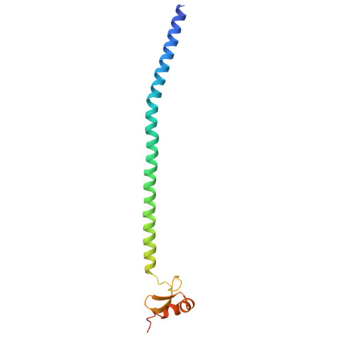 |
UniProt & NIH Common Fund Data Resources | |||||
PHAROS: Q9ULU4 GTEx: ENSG00000101040 | |||||
Entity Groups | |||||
| Sequence Clusters | 30% Identity50% Identity70% Identity90% Identity95% Identity100% Identity | ||||
| UniProt Group | Q9ULU4 | ||||
Sequence AnnotationsExpand | |||||
Reference Sequence | |||||
| Ligands 3 Unique | |||||
|---|---|---|---|---|---|
| ID | Chains | Name / Formula / InChI Key | 2D Diagram | 3D Interactions | |
| PEG (Subject of Investigation/LOI) Download:Ideal Coordinates CCD File | G [auth B], H [auth B], N [auth D] | DI(HYDROXYETHYL)ETHER C4 H10 O3 MTHSVFCYNBDYFN-UHFFFAOYSA-N |  | ||
| SO4 (Subject of Investigation/LOI) Download:Ideal Coordinates CCD File | I [auth C] | SULFATE ION O4 S QAOWNCQODCNURD-UHFFFAOYSA-L |  | ||
| ZN (Subject of Investigation/LOI) Download:Ideal Coordinates CCD File | E [auth B] F [auth B] J [auth C] K [auth C] L [auth A] | ZINC ION Zn PTFCDOFLOPIGGS-UHFFFAOYSA-N |  | ||
| Length ( Å ) | Angle ( ˚ ) |
|---|---|
| a = 63.705 | α = 90 |
| b = 71.188 | β = 90 |
| c = 230.266 | γ = 90 |
| Software Name | Purpose |
|---|---|
| PHENIX | refinement |
| XDS | data reduction |
| XSCALE | data scaling |
| PHASER | phasing |
| Funding Organization | Location | Grant Number |
|---|---|---|
| Department of Biotechnology (DBT, India) | India | BT/PR45090/MED/122/319/2022 |


