


Expression, Purification, and Structure Analysis of the Catalytic Domain of a GH167 Enzyme from Wenyingzhuangia aestuarii
Chen, F., Chang, Y.To be published.
Experimental Data Snapshot
Starting Model: in silico
View more details
wwPDB Validation 3D Report Full Report

Entity ID: 1 | |||||
|---|---|---|---|---|---|
| Molecule | Chains | Sequence Length | Organism | Details | Image |
| GH167 glycoside hydrolase | 855 | Wenyingzhuangia aestuarii | Mutation(s): 0 EC: 3.2.1 | 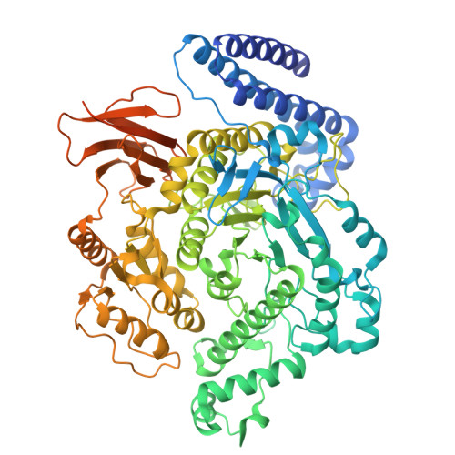 | |
| Length ( Å ) | Angle ( ˚ ) |
|---|---|
| a = 58.623 | α = 90 |
| b = 113.341 | β = 90 |
| c = 241.804 | γ = 90 |
| Software Name | Purpose |
|---|---|
| PHENIX | refinement |
| Aimless | data scaling |
| XDS | data reduction |
| PHASER | phasing |
| PDB_EXTRACT | data extraction |
| Funding Organization | Location | Grant Number |
|---|---|---|
| Other government | China | 2023YFD2100605 |


