Crystal structure of D-aspartate oxidase from Cryptococcus humicola UJ1.
Goto, M., Nonaka, R., Mizobuchi, T., Imanishi, D., Takahashi, S.(2025) Acta Crystallogr F Struct Biol Commun 81: 434-440
- PubMed: 40970329 Search on PubMed
- DOI: https://doi.org/10.1107/S2053230X25008192
- Primary Citation Related Structures:
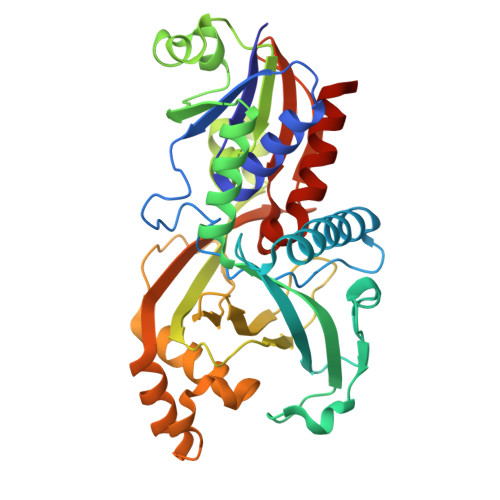
9VPF - PubMed Abstract:
The enzyme D-aspartate oxidase (DDO) oxidizes acidic D-amino acids using the coenzyme flavin adenine dinucleotide to generate the corresponding α-keto acids and ammonia. DDO differs from D-amino-acid oxidase (DAAO), which acts on neutral and basic D-amino acids. Although the enzymatic properties of DDO have been characterized in several species, the structure of DDO had remained unclear. The structure of DDO derived from Cryptococcus humicola strain UJ1 (chDDO) was determined by X-ray crystallography at 1.70 Å resolution. While the three-dimensional structures of DAAOs are known to be homodimers, chDDO forms a homotetramer. This difference was found to be caused by the deletion of one loop and the insertion of two loops.
- Faculty of Science, Toho University, 2-2-1 Miyama, Funabashi, Chiba 274-8510, Japan.
Organizational Affiliation: