Conformational Adaptability and Thermostability in alpha / beta-Peptide Fibrils Induced by beta-Amino Acid Substitution.
Li, Y., Li, D., Yao, Y., Liu, K., Zhao, Q., Zhang, Y., Xu, Y., Li, D., Sun, B., Liu, C., Dai, B.(2026) Nano Lett 26: 365-375
- PubMed: 41420871
- DOI: https://doi.org/10.1021/acs.nanolett.5c05223
- Primary Citation Related Structures:
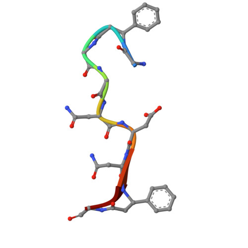
9VNK, 9VNM, 9VNN - PubMed Abstract:
The self-assembly of peptides into amyloid fibrils enables the design of functional biomaterials, yet the conformational constraints of α-peptides limit the attainable supramolecular diversity. Here, we introduce β-amino acids, β-phenylalanine (β-Phe), and β-homophenylalanine (β-hPhe) into the reversible fibril-forming core sequence hnRAC1 to generate α/β-peptide variants with distinct architectures and enhanced thermal stability. Cryo-EM reveals that β-modified peptides assemble into polymorphic fibrils with cross-β structures that differ markedly from each other and from native hnRAC1. Comparative structural analysis indicates that backbone extension by β-residues increases subunit conformational heterogeneity, enabling tighter packing and formation of more thermostable fibrils. Examination of intra- and intermolecular contacts shows that enhanced π-π stacking, hydrophobic interactions, hydrogen bonds, and electrostatic interactions likely contribute to fibril stabilization. These results show that minimal backbone modifications can remodel amyloid architecture, offering a generalizable strategy for designing structurally diverse and robust peptide-based biomaterials.
- School of Automation and Intelligent Sensing, Shanghai Jiao Tong University, Shanghai 200240, China.
Organizational Affiliation: