9VCA | pdb_00009vca

Binding site between C-reactive protein and c2cc monoclonal antibody


Experimental Data Snapshot

  • Method: ELECTRON MICROSCOPY
  • Resolution: 2.40 Å
  • Aggregation State: PARTICLE 
  • Reconstruction Method: SINGLE PARTICLE 

Starting Models: experimental
View more details

wwPDB Validation   3D Report Full Report


This is version 1.0 of the entry. See complete history


Literature

Cryo-EM structure of pentameric C-reactive protein in complex with monoclonal IgG antibodies.

Moiseenko, A.V.Kalikin, A.V.Orekhov, P.S.Byzova, N.A.Zherdev, A.V.Shaitan, K.V.Dzantiev, B.B.Sokolova, O.S.

(2025) FEBS J 

  • DOI: https://doi.org/10.1111/febs.70310
  • Primary Citation of Related Structures:  
    9VCA

  • PubMed Abstract: 

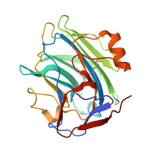
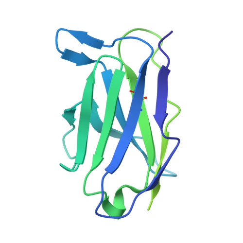
    C-reactive protein (CRP) plays a central role in innate immunity and serves as a key biomarker of inflammation. Despite its clinical importance, the structural basis of CRP interactions with antibodies remains poorly characterized. Using cryo-electron microscopy (cryo-EM), we resolved the structure of immune complexes formed between pentameric CRP and monoclonal immunoglobulin G (IgG) antibodies at up to 2.4 Å resolution. The complexes display a barrel-shaped architecture, with two CRP pentamers bridged by three to five antibodies. We built an atomic model of the CRP-antibody interface, identifying a binding site on the A-face of CRP mediated exclusively by hydrogen bonds, without salt-bridge formation. These findings provide structural insights into CRP-IgG recognition and offer a basis for the rational design of improved antibodies.


  • Organizational Affiliation
    • Faculty of Biology, Lomonosov Moscow State University, Moscow, Russia.

Macromolecules
Find similar proteins by:  (by identity cutoff)  |  3D Structure
Entity ID: 1
MoleculeChains Sequence LengthOrganismDetailsImage
C-reactive proteinA [auth E]206Homo sapiensMutation(s): 0 
Gene Names: CRPPTX1
UniProt & NIH Common Fund Data Resources
Find proteins for P02741 (Homo sapiens)
Explore P02741 
Go to UniProtKB:  P02741
PHAROS:  P02741
GTEx:  ENSG00000132693 
Entity Groups  
Sequence Clusters30% Identity50% Identity70% Identity90% Identity95% Identity100% Identity
UniProt GroupP02741
Sequence Annotations
Expand
  • Reference Sequence
Find similar proteins by:  (by identity cutoff)  |  3D Structure
Entity ID: 2
MoleculeChains Sequence LengthOrganismDetailsImage
Monoclonal IgG antibody heavy chain fragmentB [auth H]443Mus musculus albulaMutation(s): 0 
Entity Groups  
Sequence Clusters30% Identity50% Identity70% Identity90% Identity95% Identity100% Identity
Sequence Annotations
Expand
  • Reference Sequence
Find similar proteins by:  (by identity cutoff)  |  3D Structure
Entity ID: 3
MoleculeChains Sequence LengthOrganismDetailsImage
Monoclonal IgG antibody light chain fragmentC [auth L]218Mus musculus albulaMutation(s): 0 
Entity Groups  
Sequence Clusters30% Identity50% Identity70% Identity90% Identity95% Identity100% Identity
Sequence Annotations
Expand
  • Reference Sequence
Experimental Data & Validation

Experimental Data

  • Method: ELECTRON MICROSCOPY
  • Resolution: 2.40 Å
  • Aggregation State: PARTICLE 
  • Reconstruction Method: SINGLE PARTICLE 
EM Software:
TaskSoftware PackageVersion
MODEL REFINEMENTISOLDE1.8
MODEL REFINEMENTPHENIX1.20
RECONSTRUCTIONcryoSPARC4.6

Structure Validation

View Full Validation Report


Entry History & Funding Information

Deposition Data


Funding OrganizationLocationGrant Number
Not funded--

Revision History  (Full details and data files)

  • Version 1.0: 2025-11-19
    Type: Initial release