9V0L | pdb_00009v0l

UDP-binding PsBGluT, a tetrahydrobiopterin glucosyltransferase from Pseudanabaena sp. Chao 1811


Experimental Data Snapshot

  • Method: X-RAY DIFFRACTION
  • Resolution: 2.30 Å
  • R-Value Free: 
    0.264 (Depositor), 0.263 (DCC) 
  • R-Value Work: 
    0.214 (Depositor), 0.214 (DCC) 
  • R-Value Observed: 
    0.217 (Depositor) 

Starting Model: in silico
View more details

wwPDB Validation   3D Report Full Report


Ligand Structure Quality Assessment 


This is version 1.1 of the entry. See complete history


Literature

Structural analysis of the tetrahydrobiopterin glucosyltransferase PsBGluT from Pseudanabaena sp. Chao 1811.

Zang, R.Jiang, Y.Zhou, C.Z.

(2025) Acta Crystallogr F Struct Biol Commun 81: 495-504

  • DOI: https://doi.org/10.1107/S2053230X25009446
  • Primary Citation of Related Structures:  
    9V0L, 9V0W

  • PubMed Abstract: 

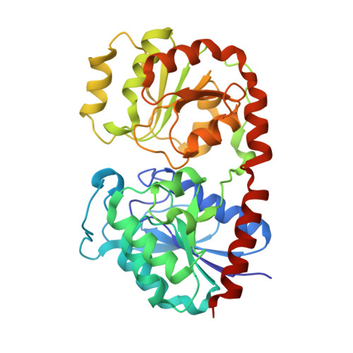
    Pterin glycosides are widely distributed in cyanobacteria and have been implicated in the regulation of phototaxis and photosynthesis. Here, we identified a new uridine diphosphate glucose:tetrahydrobiopterin α-glucosyltransferase, termed PsBGluT, from Pseudanabaena sp. Chao 1811, which catalyzes the formation of pterin glycosides. We solved crystal structures of apo PsBGluT and its UDP-bound form at 2.8 and 2.3 Å resolution, respectively. PsBGluT forms a homodimer, with each subunit adopting a canonical GT-B fold composed of two Rossmann-like domains. Structural analysis combined with molecular docking revealed the binding sites for both the donor UDP-glucose and the acceptor tetrahydrobiopterin. Based on these findings, we proposed that PsBGluT operates via an S N i retaining catalytic mechanism. This study advances our understanding of pteridine glycosylation and also provides a structural basis for investigating the photosynthetic signaling pathways in cyanobacteria.


  • Organizational Affiliation
    • Division of Life Sciences and Medicine, University of Science and Technology of China, Huang Shan Road, Hefei, Anhui 230026, People's Republic of China.

Macromolecules
Find similar proteins by:  (by identity cutoff)  |  3D Structure
Entity ID: 1
MoleculeChains Sequence LengthOrganismDetailsImage
tetrahydrobiopterin glucosyltransferases364Pseudanabaena sp. Chao 1811Mutation(s): 0 
UniProt
Find proteins for A0ABX1LL65 (Pseudanabaena yagii GIHE-NHR1)
Explore A0ABX1LL65 
Go to UniProtKB:  A0ABX1LL65
Entity Groups  
Sequence Clusters30% Identity50% Identity70% Identity90% Identity95% Identity100% Identity
UniProt GroupA0ABX1LL65
Sequence Annotations
Expand
  • Reference Sequence
Small Molecules
Ligands 1 Unique
IDChains Name / Formula / InChI Key2D Diagram3D Interactions
UDP (Subject of Investigation/LOI)
Query on UDP

Download Ideal Coordinates CCD File 
B [auth A]URIDINE-5'-DIPHOSPHATE
C9 H14 N2 O12 P2
XCCTYIAWTASOJW-XVFCMESISA-N
Experimental Data & Validation

Experimental Data

  • Method: X-RAY DIFFRACTION
  • Resolution: 2.30 Å
  • R-Value Free:  0.264 (Depositor), 0.263 (DCC) 
  • R-Value Work:  0.214 (Depositor), 0.214 (DCC) 
  • R-Value Observed: 0.217 (Depositor) 
Space Group: P 61 2 2
Unit Cell:
Length ( Å )Angle ( ˚ )
a = 176.97α = 90
b = 176.97β = 90
c = 66.171γ = 120
Software Package:
Software NamePurpose
PHENIXrefinement
CrysalisProdata reduction
Aimlessdata scaling
MOLREPphasing

Structure Validation

View Full Validation Report


Ligand Structure Quality Assessment 


Entry History & Funding Information

Deposition Data


Funding OrganizationLocationGrant Number
National Natural Science Foundation of China (NSFC)China32430001

Revision History  (Full details and data files)

  • Version 1.0: 2025-11-26
    Type: Initial release
  • Version 1.1: 2025-12-17
    Changes: Database references