9UBQ | pdb_00009ubq

HBsAg in complex with H020 Fab


Experimental Data Snapshot

  • Method: ELECTRON MICROSCOPY
  • Resolution: 2.86 Å
  • Aggregation State: PARTICLE 
  • Reconstruction Method: SINGLE PARTICLE 

wwPDB Validation   3D Report Full Report


This is version 1.1 of the entry. See complete history


Literature

The Symmetric Structure of the Antigenic Loop in Type B HBV Surface Antigen.

Tao, W.He, X.Chen, L.

(2025) J Mol Biology 437: 169483-169483

  • DOI: https://doi.org/10.1016/j.jmb.2025.169483
  • Primary Citation of Related Structures:  
    9UBQ

  • PubMed Abstract: 

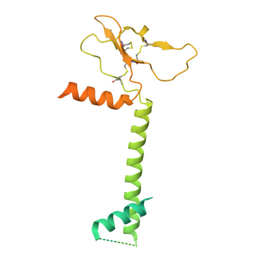
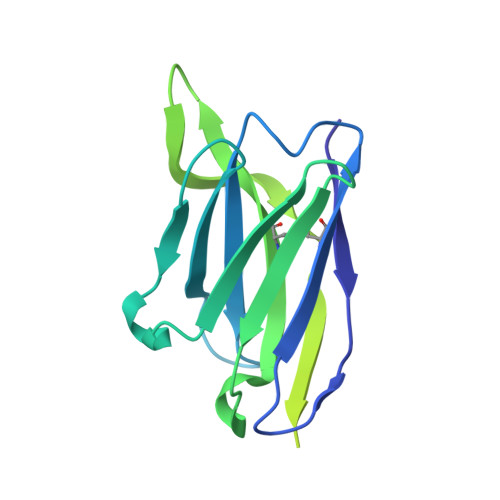
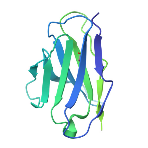
    Hepatitis B virus (HBV) is an enveloped virus with HBV surface antigen (HBsAg) as the only protein on its viral membrane. The extracellular antigenic loop (AGL) of HBsAg plays a crucial role in viral attachment to host cells, serves as the primary target for neutralizing antibodies (NAbs), and is subject to escape mutations. Previous studies have shown that the AGL exhibits two different structures (Type A and Type B) dictated by distinct disulfide bond linkage. However, due to the flexibility of some regions in previous structure, the complete model of AGL Type B and its symmetry remain elusive. Here, we present the cryo-EM structure of AGL Type B in complex with the Fab fragment of the NAb H020. The complete structure of AGL Type B reveals its two-fold symmetry and it can bind two Fab H020 fragments simultaneously. Further analysis elucidates the underlying mechanism of pan-serotype neutralizing capability of H020 and how escape mutations hinder its binding.


  • Organizational Affiliation
    • State Key Laboratory of Membrane Biology, College of Future Technology, Institute of Molecular Medicine, Peking. University, Beijing Key Laboratory of Cardiometabolic Molecular Medicine, Beijing 100871, China.

Macromolecules
Find similar proteins by:  (by identity cutoff)  |  3D Structure
Entity ID: 1
MoleculeChains Sequence LengthOrganismDetailsImage
Middle S proteinA,
D [auth B]
281Hepatitis B virusMutation(s): 3 
UniProt
Find proteins for B5TFB1 (Hepatitis B virus)
Explore B5TFB1 
Go to UniProtKB:  B5TFB1
Entity Groups  
Sequence Clusters30% Identity50% Identity70% Identity90% Identity95% Identity100% Identity
UniProt GroupB5TFB1
Sequence Annotations
Expand
  • Reference Sequence
Find similar proteins by:  (by identity cutoff)  |  3D Structure
Entity ID: 2
MoleculeChains Sequence LengthOrganismDetailsImage
H020 Fab Heavy ChainB [auth C],
E
220Homo sapiensMutation(s): 0 
Entity Groups  
Sequence Clusters30% Identity50% Identity70% Identity90% Identity95% Identity100% Identity
Sequence Annotations
Expand
  • Reference Sequence
Find similar proteins by:  (by identity cutoff)  |  3D Structure
Entity ID: 3
MoleculeChains Sequence LengthOrganismDetailsImage
H020 Fab Light ChainC [auth D],
F
215Homo sapiensMutation(s): 0 
Entity Groups  
Sequence Clusters30% Identity50% Identity70% Identity90% Identity95% Identity100% Identity
Sequence Annotations
Expand
  • Reference Sequence
Experimental Data & Validation

Experimental Data

  • Method: ELECTRON MICROSCOPY
  • Resolution: 2.86 Å
  • Aggregation State: PARTICLE 
  • Reconstruction Method: SINGLE PARTICLE 

Structure Validation

View Full Validation Report


Entry History & Funding Information

Deposition Data


Funding OrganizationLocationGrant Number
National Natural Science Foundation of China (NSFC)China31821091

Revision History  (Full details and data files)

  • Version 1.0: 2025-10-29
    Type: Initial release
  • Version 1.1: 2025-11-05
    Changes: Data collection, Database references