Structural Mimicry Without Glyoxalase I Functional Convergence: A Homogentisate 1,2-Dioxygenase From Acinetobacter.
Seo, P.W., Hwangbo, S.A., Kim, J.S., Park, S.Y.(2025) Proteins 93: 2150-2157
- PubMed: 40650421 Search on PubMed
- DOI: https://doi.org/10.1002/prot.70020
- Primary Citation Related Structures:
9U42 - PubMed Abstract:
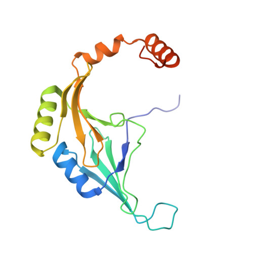
Homogentisate 1,2-dioxygenase (HGD) is a non-heme iron enzyme that plays a crucial role in phenylalanine and tyrosine metabolism. Acinetobacter-derived HGD (AcHGD) exhibits structural similarity to glyoxalase I (GLO1) but lacks GLO1 activity. In this study, we analyzed the crystal structure of AcHGD at a resolution of 1.5 Å and investigated the molecular basis for its lack of GLO1 activity using enzymatic assays, isothermal titration calorimetry (ITC), and site-directed mutagenesis. Metal ion dependency assays revealed that AcHGD exhibits high specificity for Fe 2+ , supporting its role as a non-heme iron (II)-dependent dioxygenase. Structural analysis revealed that AcHGD adopts a β-barrel fold similar to GLO1 and coordinates Zn 2+ through a 2-His-1-carboxylate facial triad. However, its substrate-binding tunnel is narrower than that of GLO1, preventing the binding of S-D-lactoylglutathione, the natural substrate of GLO1. Moreover, introducing GLO1-like mutations in the active site failed to confer GLO1 activity and instead abolished HGD activity. ITC analysis confirmed that AcHGD binds strongly to homogentisate but does not interact with S-D-lactoylglutathione. These findings demonstrate that despite its structural resemblance to GLO1, AcHGD lacks GLO1 activity due to differences in substrate specificity and active site architecture. This study provides insights into the structure-function relationship and evolutionary divergence between HGD and GLO1 enzymes.
- Department of Life Sciences, Pohang University of Science and Technology, Pohang, Gyeongbuk, Korea.
Organizational Affiliation: