9RTL | pdb_00009rtl

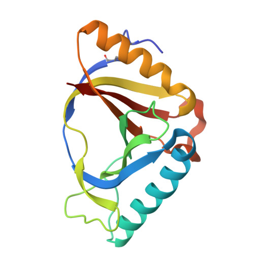
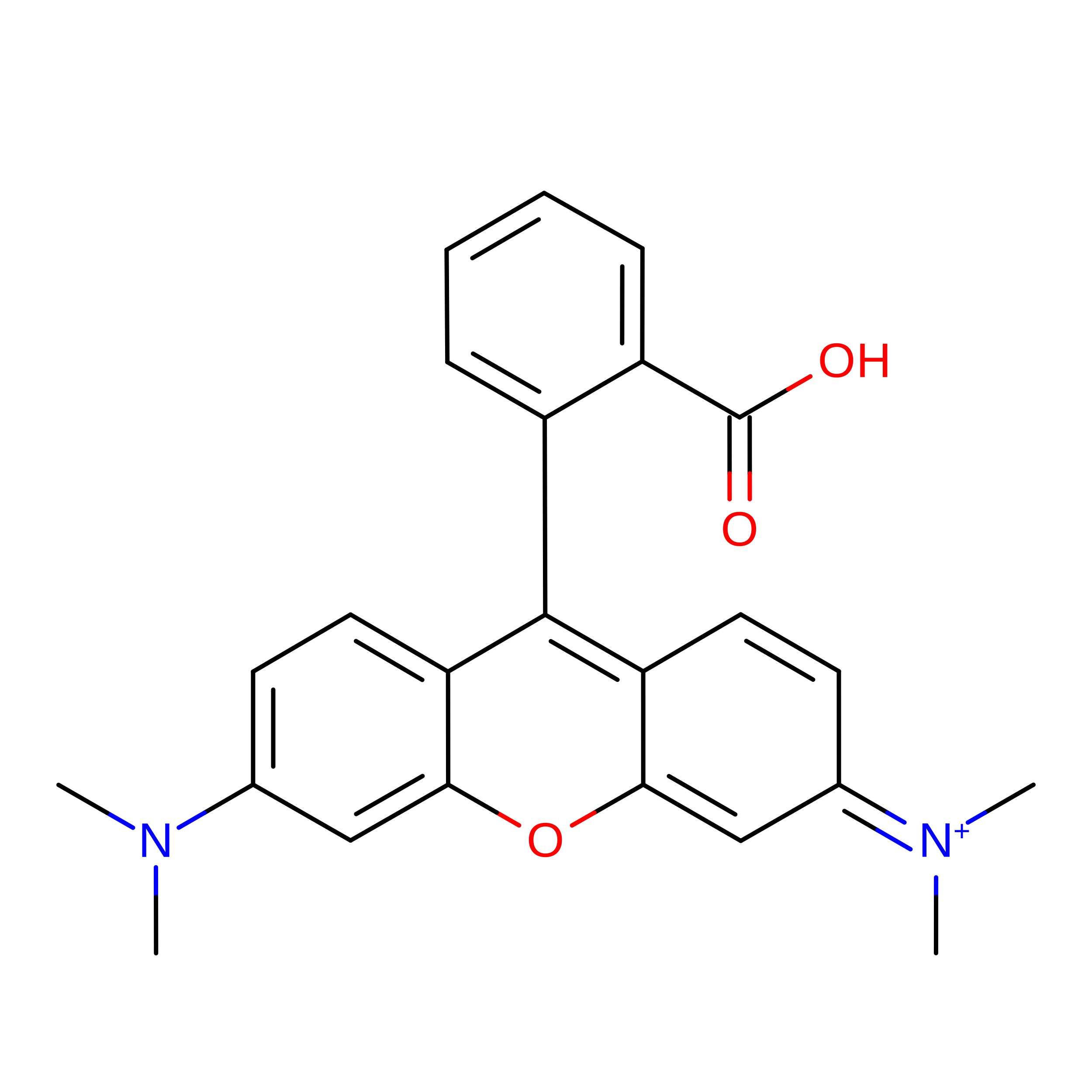
X-ray structure of the CTR107-N138A mutant bound to a tetramethylrhodamine ligand


Experimental Data Snapshot

  • Method: X-RAY DIFFRACTION
  • Resolution: 1.80 Å
  • R-Value Free: 
    0.213 (Depositor), 0.213 (DCC) 
  • R-Value Work: 
    0.182 (Depositor), 0.182 (DCC) 
  • R-Value Observed: 
    0.184 (Depositor) 

Starting Model: experimental
View more details

wwPDB Validation 3D Report Full Report

Validation slider image for 9RTL

Ligand Structure Quality Assessment 


This is version 1.1 of the entry. See complete history

Literature

Fast, Bright, and Reversible Fluorescent Labeling of Rhodamine-Binding Proteins.

Kompa, J.Dornfeld, L.J.Porzberg, N.Jang, S.Zedlitz, S.Lilje, S.H.Catapano, C.Jocher, D.Merk, L.Hoege, C.Mao, R.Wilhelm, J.Dietz, M.S.Tarnawski, M.Hiblot, J.Hyman, A.A.Heilemann, M.Johnsson, K.

(2026) J Am Chem Soc 148: 1419-1429

  • DOI: https://doi.org/10.1021/jacs.5c18083
  • Primary Citation Related Structures: 
    9RTL, 9RTM

  • PubMed Abstract: 

    Rhodamine dyes conjugated to targeting ligands can yield exceptionally bright fluorescent probes for live-cell imaging. However, the limited permeability of such rhodamine derivatives restricts their broader applications, particularly in vivo . Here, we present Rho-tag and SiR-tag, engineered protein tags derived from bacterial multidrug-resistant proteins that bind unsubstituted (silicon) rhodamines with nanomolar affinity. Unsubstituted (silicon) rhodamines readily cross membranes and enable rapid, reversible, and fluorogenic labeling of the tags in mammalian cells within seconds. The labeling of Rho-tag and SiR-tag is compatible with various super-resolution imaging methods and allows their use alongside self-labeling tags, such as HaloTag7 and SNAP-tag. The high affinity and specificity of both tags, combined with the permeability and outstanding spectroscopic properties of rhodamines, make them particularly attractive for in vivo bioimaging, as demonstrated by efficient fluorescent labeling in C. elegans embryos and zebrafish larvae.


  • Organizational Affiliation
    • Department of Chemical Biology, Max Planck Institute for Medical Research, Heidelberg 69120, Germany.

Macromolecule Content 

  • Total Structure Weight: 35.82 kDa 
  • Atom Count: 2,554 
  • Modeled Residue Count: 309 
  • Deposited Residue Count: 320 
  • Unique protein chains: 1

Macromolecules

Find similar proteins by:|  3D Structure
Entity ID: 1
MoleculeChains  Sequence LengthOrganismDetailsImage
AraC effector-binding domain-containing protein
A, B
160Chlorobaculum tepidum TLSMutation(s): 1 
Gene Names: CT0179
UniProt
Find proteins for Q8KFZ1 (Chlorobaculum tepidum (strain ATCC 49652 / DSM 12025 / NBRC 103806 / TLS))
Explore Q8KFZ1 
Go to UniProtKB:  Q8KFZ1
Entity Groups
Sequence Clusters30% Identity50% Identity70% Identity90% Identity95% Identity100% Identity
UniProt GroupQ8KFZ1
Sequence Annotations
Expand
Reference Sequence

Small Molecules

Experimental Data & Validation

Experimental Data

  • Method: X-RAY DIFFRACTION
  • Resolution: 1.80 Å
  • R-Value Free:  0.213 (Depositor), 0.213 (DCC) 
  • R-Value Work:  0.182 (Depositor), 0.182 (DCC) 
  • R-Value Observed: 0.184 (Depositor) 
Space Group: P 65
Unit Cell:
Length ( Å )Angle ( ˚ )
a = 71.81α = 90
b = 71.81β = 90
c = 146.16γ = 120
Software Package:
Software NamePurpose
PHENIXrefinement
XDSdata reduction
XSCALEdata scaling
PHASERphasing

Structure Validation

View Full Validation Report


Ligand Structure Quality Assessment 


Entry History 

& Funding Information

Deposition Data


Funding OrganizationLocationGrant Number
Max Planck SocietyGermany--

Revision History  (Full details and data files)

  • Version 1.0: 2025-07-30
    Type: Initial release
  • Version 1.1: 2026-08-26
    Changes: Database references