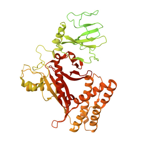
Molecular basis for anti-jumbo phage immunity by AVAST type 5.
Muralidharan, A., Costa, A.R., Fierlier, D., van den Berg, D.F., van den Bossche, H., Zoumaro-Djayoon, A.D., Rodriguez-Molina, A., Pabst, M., Pacesa, M., Correia, B.E., Brouns, S.J.J.(2026) Mol Cell 86: 740
- PubMed: 41653918 Search on PubMed
- DOI: https://doi.org/10.1016/j.molcel.2026.01.004
- Primary Citation Related Structures:
9RP3 - PubMed Abstract:
Jumbo phages protect their genomes from DNA-sensing bacterial defense systems by enclosing them within vesicles and nucleus-like compartments. Very little is known about defense systems specialized to counter these phages. Here, we show that AVAST type 5 (Avs5) systems, part of the signal transduction ATPases of numerous domains (STAND) superfamily, confer conserved immunity against jumbo phages. Using fluorescence microscopy and biotin proximity labeling, we demonstrate that Avs5 localizes to early infection vesicles, where it senses an essential, early-expressed phage protein named JADA (Jumbo phage Avs5 Defense Activator). Recognition of phage infection triggers the Sir2-like effector domain of Avs5 across three Avs5 clades, resulting in rapid NAD + hydrolysis, disruption of phage nucleus formation, and arrest of infection. These findings reveal a spatially coordinated bacterial immune strategy that targets an early vulnerability in jumbo phage infection.
- Department of Bionanoscience, Delft University of Technology, van der Maasweg 9, 2629 HZ Delft, the Netherlands; Kavli Institute of Nanoscience, Delft University of Technology, van der Maasweg 9, 2629 HZ Delft, the Netherlands.
Organizational Affiliation: