9RAT | pdb_00009rat

EFFECTS OF TEMPERATURE ON PROTEIN STRUCTURE AND DYNAMICS: X-RAY CRYSTALLOGRAPHIC STUDIES OF THE PROTEIN RIBONUCLEASE-A AT NINE DIFFERENT TEMPERATURES FROM 98 TO 320 K


Experimental Data Snapshot

  • Method: X-RAY DIFFRACTION
  • Resolution: 1.50 Å
  • R-Value Observed: 
    0.191 (Depositor) 

wwPDB Validation 3D Report Full Report

Validation slider image for 9RAT

This is version 1.4 of the entry. See complete history

Literature

Effects of temperature on protein structure and dynamics: X-ray crystallographic studies of the protein ribonuclease-A at nine different temperatures from 98 to 320 K.

Tilton Jr., R.F.Dewan, J.C.Petsko, G.A.

(1992) Biochemistry 31: 2469-2481

  • DOI: https://doi.org/10.1021/bi00124a006
  • Primary Citation Related Structures: 
    1RAT, 2RAT, 3RAT, 4RAT, 5RAT, 6RAT, 7RAT, 8RAT, 9RAT

  • PubMed Abstract: 

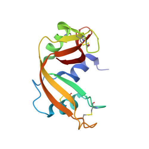
    Structures using X-ray diffraction data collected to 1.5-A resolution have been determined for the protein ribonuclease-A at nine different temperatures ranging from 98 to 320 K. It is determined that the protein molecule expands slightly (0.4% per 100 K) with increasing temperature and that this expansion is linear. The expansion is due primarily to subtle repacking of the molecule, with exposed and mobile loop regions exhibiting the largest movements. Individual atomic Debye-Waller factors exhibit predominantly biphasic behavior, with a small positive slope at low temperatures and a larger positive slope at higher temperatures. The break in this curve occurs at a characteristic temperature of 180-200 K, perhaps indicative of fundamental changes in the dynamical structure of the surrounding protein solvent. The distribution of protein Debye-Waller factors is observed to broaden as well as shift to higher values as the temperature is increased.


  • Organizational Affiliation
    • Miles Research Center, Miles Inc., West Haven, Connecticut 06516.

Macromolecule Content 

  • Total Structure Weight: 13.71 kDa 
  • Atom Count: 951 
  • Modeled Residue Count: 124 
  • Deposited Residue Count: 124 
  • Unique protein chains: 1

Macromolecules

Find similar proteins by:|  3D Structure
Entity ID: 1
MoleculeChains  Sequence LengthOrganismDetailsImage
RIBONUCLEASE A124Bos taurusMutation(s): 0 
EC: 3.1.27.5 (PDB Primary Data), 4.6.1.18 (UniProt)
UniProt
Find proteins for P61823 (Bos taurus)
Explore P61823 
Go to UniProtKB:  P61823
Entity Groups
Sequence Clusters30% Identity50% Identity70% Identity90% Identity95% Identity100% Identity
UniProt GroupP61823
Sequence Annotations
Expand
Reference Sequence

Experimental Data & Validation

Experimental Data

  • Method: X-RAY DIFFRACTION
  • Resolution: 1.50 Å
  • R-Value Observed: 0.191 (Depositor) 
Space Group: P 1 21 1
Unit Cell:
Length ( Å )Angle ( ˚ )
a = 30.4α = 90
b = 38.3β = 106.9
c = 53.3γ = 90
Software Package:
Software NamePurpose
PROLSQrefinement

Structure Validation

View Full Validation Report


Entry History 

Deposition Data

Revision History  (Full details and data files)

  • Version 1.0: 1993-07-15
    Type: Initial release
  • Version 1.1: 2008-03-25
    Changes: Version format compliance
  • Version 1.2: 2011-07-13
    Changes: Version format compliance
  • Version 1.3: 2024-06-05
    Changes: Data collection, Database references, Other
  • Version 1.4: 2024-10-23
    Changes: Structure summary