9R35 | pdb_00009r35

Crystal structure of the Pseudomonas putida Xre-RES toxin-antitoxin complex bound to promoter DNA


Experimental Data Snapshot

  • Method: X-RAY DIFFRACTION
  • Resolution: 2.70 Å
  • R-Value Free: 
    0.269 (Depositor), 0.272 (DCC) 
  • R-Value Work: 
    0.228 (Depositor), 0.245 (DCC) 

Starting Model: experimental
View more details

wwPDB Validation   3D Report Full Report


This is version 1.1 of the entry. See complete history


Literature

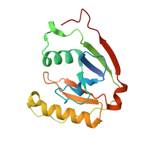
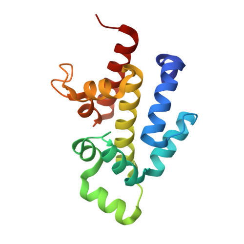
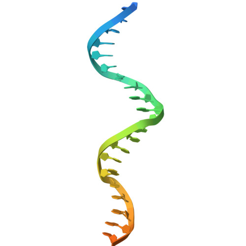
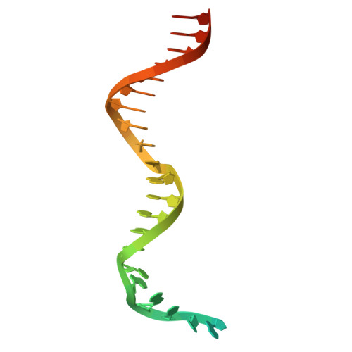
Structural basis for higher-order DNA binding by a bacterial transcriptional regulator.

Henriksen, F.O.G.Van, L.B.Brodersen, D.E.Skjerning, R.B.

(2025) PLoS Genet 21: e1011749-e1011749

  • DOI: https://doi.org/10.1371/journal.pgen.1011749
  • Primary Citation of Related Structures:  
    9R35

  • PubMed Abstract: 

    Transcriptional regulation by binding of transcription factors to palindromic sequences in promoter regions is a fundamental process in bacteria. Some transcription factors have multiple dimeric DNA-binding domains, in principle enabling interaction with higher-order DNA structures; however, mechanistic and structural insights into this phenomenon remain limited. The Pseudomonas putida toxin-antitoxin (TA) system Xre-RES has an unusual 4:2 stoichiometry including two potential DNA-binding sites, compatible with a complex mechanism of transcriptional autoregulation. Here, we show that the Xre-RES complex interacts specifically with a palindromic DNA repeat in the promoter in a 1:1 molar ratio, leading to transcriptional repression. We determine the 2.7 Å crystal structure of the protein-DNA complex, revealing an unexpected asymmetry in the interaction and suggesting the presence of a secondary binding site, which is supported by structural prediction of the binding to the intact promoter region. Additionally, we show that the antitoxin can be partially dislodged from the Xre-RES complex, resulting in Xre monomers and a 2:2 Xre-RES complex, neither of which repress transcription. These findings highlight a dynamic, concentration-dependent model of transcriptional autoregulation, in which the Xre-RES complex transitions between a non-binding (2:2) and a DNA-binding (4:2) form.


  • Organizational Affiliation
    • Department of Molecular Biology and Genetics, Aarhus University, Aarhus, Denmark.

Macromolecules

Find similar proteins by:  (by identity cutoff)  |  3D Structure
Entity ID: 1
MoleculeChains Sequence LengthOrganismDetailsImage
Toxin Res
A, D, G, J, M
A, D, G, J, M, P, S, V
158Pseudomonas putida KT2440Mutation(s): 0 
Gene Names: resPP_2434
EC: 2.4.2
UniProt
Find proteins for Q88K57 (Pseudomonas putida (strain ATCC 47054 / DSM 6125 / CFBP 8728 / NCIMB 11950 / KT2440))
Explore Q88K57 
Go to UniProtKB:  Q88K57
Entity Groups  
Sequence Clusters30% Identity50% Identity70% Identity90% Identity95% Identity100% Identity
UniProt GroupQ88K57
Sequence Annotations
Expand
  • Reference Sequence
Find similar proteins by:  (by identity cutoff)  |  3D Structure
Entity ID: 2
MoleculeChains Sequence LengthOrganismDetailsImage
XRE anti-toxin
B, C, E, F, H
B, C, E, F, H, I, K, L, N, O, Q, R, T, U, W, X
149Pseudomonas putida KT2440Mutation(s): 0 
Gene Names: AYO08_12380B7H19_10555CBL13_00496CBP06_13110
UniProt
Find proteins for A0A179RFM7 (Pseudomonas putida)
Explore A0A179RFM7 
Go to UniProtKB:  A0A179RFM7
Entity Groups  
Sequence Clusters30% Identity50% Identity70% Identity90% Identity95% Identity100% Identity
UniProt GroupA0A179RFM7
Sequence Annotations
Expand
  • Reference Sequence
Find similar nucleic acids by:  (by identity cutoff)  |  3D Structure
Entity ID: 3
MoleculeChains LengthOrganismImage
DNA reverse (30-mer)BA [auth d],
DA [auth f],
FA [auth h],
Y [auth a]
30Pseudomonas putida KT2440
Sequence Annotations
Expand
  • Reference Sequence
Find similar nucleic acids by:  (by identity cutoff)  |  3D Structure
Entity ID: 4
MoleculeChains LengthOrganismImage
DNA forward (30-mer)AA [auth c],
CA [auth e],
EA [auth g],
Z [auth b]
30Pseudomonas putida KT2440
Sequence Annotations
Expand
  • Reference Sequence
Experimental Data & Validation

Experimental Data

  • Method: X-RAY DIFFRACTION
  • Resolution: 2.70 Å
  • R-Value Free:  0.269 (Depositor), 0.272 (DCC) 
  • R-Value Work:  0.228 (Depositor), 0.245 (DCC) 
Space Group: P 1 21 1
Unit Cell:
Length ( Å )Angle ( ˚ )
a = 81.23α = 90
b = 185.11β = 90.01
c = 170.79γ = 90
Software Package:
Software NamePurpose
PHENIXrefinement
XDSdata reduction
XSCALEdata scaling
PHASERphasing
PDB_EXTRACTdata extraction

Structure Validation

View Full Validation Report


Entry History & Funding Information

Deposition Data


Funding OrganizationLocationGrant Number
Novo Nordisk FoundationDenmarkNNF18OC0030646
Novo Nordisk FoundationDenmarkNNF22OC0079855
Independent Research Fund Denmark - Medical SciencesDenmark0135-00072B

Revision History  (Full details and data files)

  • Version 1.0: 2025-08-20
    Type: Initial release
  • Version 1.1: 2025-09-03
    Changes: Database references