9QT1 | pdb_00009qt1

Bacterial histone HLp from Leptospira perolatii bound to DNA


Experimental Data Snapshot

  • Method: X-RAY DIFFRACTION
  • Resolution: 2.10 Å
  • R-Value Free: 
    0.279 (Depositor), 0.296 (DCC) 
  • R-Value Work: 
    0.250 (Depositor), 0.261 (DCC) 
  • R-Value Observed: 
    0.251 (Depositor) 

Starting Model: experimental
View more details

wwPDB Validation   3D Report Full Report


This is version 1.0 of the entry. See complete history


Literature

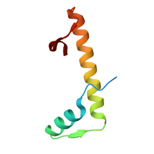
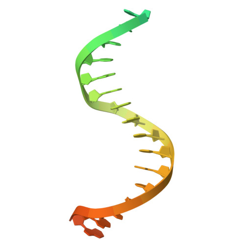
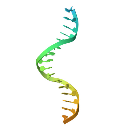
DNA Wrapping by a tetrameric bacterial histone.

Hu, Y.Schwab, S.Qiu, K.Zhang, Y.Bar, K.Reichle, H.Panzera, A.Lupas, A.N.Hartmann, M.D.Dame, R.T.Alva, V.Hernandez Alvarez, B.

(2025) Nat Commun 16: 11108-11108

  • DOI: https://doi.org/10.1038/s41467-025-67425-w
  • Primary Citation of Related Structures:  
    9QT0, 9QT1, 9QT2

  • PubMed Abstract: 

    Histones are conserved DNA-packaging proteins found across all domains of life. In eukaryotes, canonical histones form octamers that wrap ~147 base pairs (bp) of DNA into nucleosomes, while in archaea they form dimers that polymerize into extended hypernucleosomes. Although bacteria were long thought to lack histones, homologs have now been identified in diverse lineages. We previously characterized the histone HBb from Bdellovibrio bacteriovorus, which binds and bends DNA as a dimer. Here, we describe HLp from Leptospira perolatii and show by crystallographic and biophysical analyses that, unlike HBb, it forms stable tetramers and binds DNA nonspecifically, wrapping ~60 bp of DNA around its core. Molecular dynamics simulations, DNA-binding assays, and heterologous expression in Escherichia coli, where HLp reorganizes the nucleoid, support a role in bacterial chromatin organization. These findings expand the repertoire of bacterial histone-DNA interactions and highlight the diversity of histone-based genome organization across the tree of life.


  • Organizational Affiliation
    • Department of Protein Evolution, Max Planck Institute for Biology Tübingen, Tübingen, Germany.

Macromolecules

Find similar proteins by:  (by identity cutoff)  |  3D Structure
Entity ID: 1
MoleculeChains Sequence LengthOrganismDetailsImage
DUF1931 domain-containing protein
A, B
65Leptospira perolatiiMutation(s): 0 
Gene Names: CH373_10155
UniProt
Find proteins for A0A2M9ZN55 (Leptospira perolatii)
Explore A0A2M9ZN55 
Go to UniProtKB:  A0A2M9ZN55
Entity Groups  
Sequence Clusters30% Identity50% Identity70% Identity90% Identity95% Identity100% Identity
UniProt GroupA0A2M9ZN55
Sequence Annotations
Expand
  • Reference Sequence
Find similar nucleic acids by:  (by identity cutoff)  |  3D Structure
Entity ID: 2
MoleculeChains LengthOrganismImage
DNA30DNA molecule
Sequence Annotations
Expand
  • Reference Sequence
Find similar nucleic acids by:  (by identity cutoff)  |  3D Structure
Entity ID: 3
MoleculeChains LengthOrganismImage
DNA30DNA molecule
Sequence Annotations
Expand
  • Reference Sequence
Experimental Data & Validation

Experimental Data

  • Method: X-RAY DIFFRACTION
  • Resolution: 2.10 Å
  • R-Value Free:  0.279 (Depositor), 0.296 (DCC) 
  • R-Value Work:  0.250 (Depositor), 0.261 (DCC) 
  • R-Value Observed: 0.251 (Depositor) 
Space Group: P 41 21 2
Unit Cell:
Length ( Å )Angle ( ˚ )
a = 67.934α = 90
b = 67.934β = 90
c = 97.902γ = 90
Software Package:
Software NamePurpose
XDSdata reduction
XDSdata scaling
MOLREPphasing
REFMACrefinement

Structure Validation

View Full Validation Report


Entry History & Funding Information

Deposition Data


Funding OrganizationLocationGrant Number
Max Planck SocietyGermany--

Revision History  (Full details and data files)

  • Version 1.0: 2025-12-24
    Type: Initial release