Impurities in amyloid studies: The power of automated model building within a cautionary tale for structural biologists.
Rhyner, D., Frey, L., Zhou, J., Kwiatkowski, W., Mezzenga, R., Riek, R., Greenwald, J.(2025) Protein Sci 34: e70353-e70353
- PubMed: 41123416 Search on PubMedSearch on PubMed Central
- DOI: https://doi.org/10.1002/pro.70353
- Primary Citation Related Structures:
9QLU - PubMed Abstract:
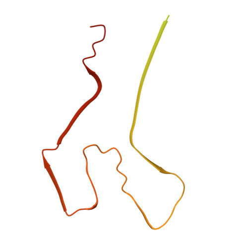
The purity of protein samples of biological origin is often difficult to ascertain, leading the naïve or optimistic scientist to underestimate contaminants in their research. Even after extensive purification, protein samples can contain nucleic acids, truncated degradation products, or other protein contaminants. While in many cases, and when present at low concentrations, such contaminants are unlikely to alter experimental results significantly, they must be considered when studying protein aggregation. Such reactions can be sensitive to small environmental changes in their early stages due to a nucleation-dependent mechanism, where minor differences can be amplified during the subsequent exponential growth phase. During a recent study of the amyloid formation of human lysozyme, we encountered a significant amyloid-forming protein contaminant derived from the expression host Oryza sativa japonica. Further investigation of this widely used commercial source of human lysozyme revealed at least a dozen protein contaminants. These discoveries led to intriguing observations, including an underdeveloped branch of plant amyloid research and a possible link between the amyloid fold and allergens. Here, we present our findings within a cautionary tale for structural biologists: a surprising variety of contaminants in a commercial protein sample and the accidental yet definitive identification of one of them by cryo-electron microscopy helical reconstruction. The resulting 2.54 Å model of the 17 kDa alpha-amylase/trypsin inhibitor Type 2 marks the first known amyloid structure of a plant protein.
- Institute of Molecular Physical Science, ETH Zürich, Zürich, Switzerland.
Organizational Affiliation: