9QFQ | pdb_00009qfq

Cryo-EM structure of the cofilactin barbed end bound by AIP1


Experimental Data Snapshot

  • Method: ELECTRON MICROSCOPY
  • Resolution: 2.76 Å
  • Aggregation State: FILAMENT 
  • Reconstruction Method: SINGLE PARTICLE 

Starting Models: experimental, in silico
View more details

wwPDB Validation   3D Report Full Report


This is version 1.2 of the entry. See complete history


Literature

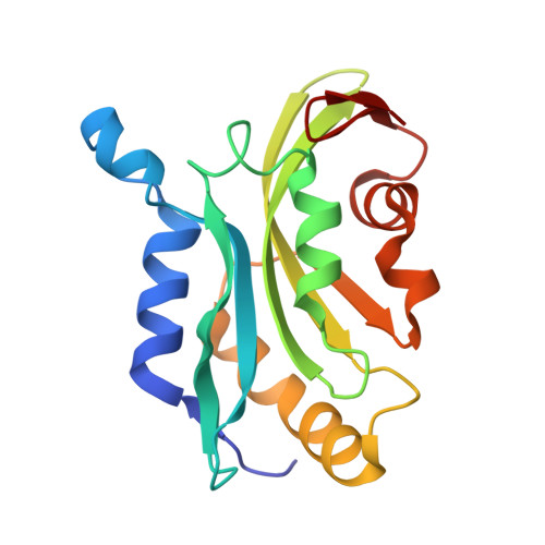
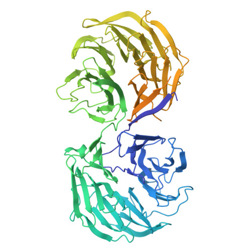
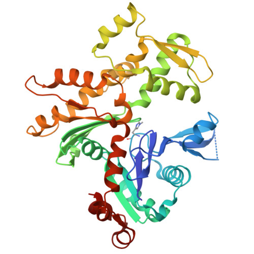
Choreography of rapid actin filament disassembly by coronin, cofilin, and AIP1.

Oosterheert, W.Boiero Sanders, M.Hofnagel, O.Bieling, P.Raunser, S.

(2025) Cell 188: 6845

  • DOI: https://doi.org/10.1016/j.cell.2025.09.016
  • Primary Citation of Related Structures:  
    9QEW, 9QEY, 9QF2, 9QFB, 9QFD, 9QFE, 9QFG, 9QFJ, 9QFK, 9QFO, 9QFQ, 9QFW

  • PubMed Abstract: 

    Rapid remodeling of actin filament (F-actin) networks is essential for the movement and morphogenesis of eukaryotic cells. The conserved actin-binding proteins coronin, cofilin, and actin-interacting protein 1 (AIP1) act in synergy to promote rapid F-actin network disassembly, but the underlying mechanisms have remained elusive. Here, using cryo-electron microscopy (cryo-EM), we uncover the concerted molecular actions of coronin, cofilin, and AIP1 that lead to actin filament aging and severing. We find that the cooperative binding of coronin allosterically promotes inorganic phosphate release from F-actin and induces filament undertwisting, thereby priming the filament for cofilin binding. Cofilin then displaces coronin from the filament via a strand-restricted cooperative binding mechanism. The resulting cofilactin serves as a high-affinity platform for AIP1, which induces severing by acting as a clamp that disrupts inter-subunit filament contacts. In this "molecular squeezing" mechanism, AIP1 and not cofilin is responsible for filament severing. Our work redefines the role of key disassembly factors in actin dynamics.


  • Organizational Affiliation
    • Department of Structural Biochemistry, Max Planck Institute of Molecular Physiology, 44227 Dortmund, Germany. Electronic address: w.oosterheert@nki.nl.

Macromolecules
Find similar proteins by:  (by identity cutoff)  |  3D Structure
Entity ID: 1
MoleculeChains Sequence LengthOrganismDetailsImage
Cofilin-1A [auth F],
B [auth G],
C [auth H],
D [auth I],
E [auth J]
166Homo sapiensMutation(s): 0 
Gene Names: CFL1CFL
UniProt & NIH Common Fund Data Resources
Find proteins for P23528 (Homo sapiens)
Explore P23528 
Go to UniProtKB:  P23528
PHAROS:  P23528
GTEx:  ENSG00000172757 
Entity Groups  
Sequence Clusters30% Identity50% Identity70% Identity90% Identity95% Identity100% Identity
UniProt GroupP23528
Sequence Annotations
Expand
  • Reference Sequence
Find similar proteins by:  (by identity cutoff)  |  3D Structure
Entity ID: 2
MoleculeChains Sequence LengthOrganismDetailsImage
WD repeat-containing protein 1,Methylated-DNA--protein-cysteine methyltransferaseF [auth P]805Homo sapiensMutation(s): 0 
Gene Names: WDR1MGMT
EC: 2.1.1.63
UniProt & NIH Common Fund Data Resources
Find proteins for O75083 (Homo sapiens)
Explore O75083 
Go to UniProtKB:  O75083
PHAROS:  O75083
GTEx:  ENSG00000071127 
Find proteins for P16455 (Homo sapiens)
Explore P16455 
Go to UniProtKB:  P16455
PHAROS:  P16455
GTEx:  ENSG00000170430 
Entity Groups  
Sequence Clusters30% Identity50% Identity70% Identity90% Identity95% Identity100% Identity
UniProt GroupsP16455O75083
Sequence Annotations
Expand
  • Reference Sequence
Find similar proteins by:  (by identity cutoff)  |  3D Structure
Entity ID: 3
MoleculeChains Sequence LengthOrganismDetailsImage
Actin, cytoplasmic 1, N-terminally processedG [auth A],
H [auth B],
I [auth C],
J [auth D],
K [auth E]
374Homo sapiensMutation(s): 1 
Gene Names: ACTB
EC: 3.6.4
UniProt & NIH Common Fund Data Resources
Find proteins for P60709 (Homo sapiens)
Explore P60709 
Go to UniProtKB:  P60709
PHAROS:  P60709
GTEx:  ENSG00000075624 
Entity Groups  
Sequence Clusters30% Identity50% Identity70% Identity90% Identity95% Identity100% Identity
UniProt GroupP60709
Sequence Annotations
Expand
  • Reference Sequence
Small Molecules
Ligands 2 Unique
IDChains Name / Formula / InChI Key2D Diagram3D Interactions
ADP (Subject of Investigation/LOI)
Query on ADP

Download Ideal Coordinates CCD File 
M [auth A],
O [auth B],
Q [auth C],
S [auth D],
U [auth E]
ADENOSINE-5'-DIPHOSPHATE
C10 H15 N5 O10 P2
XTWYTFMLZFPYCI-KQYNXXCUSA-N
MG (Subject of Investigation/LOI)
Query on MG

Download Ideal Coordinates CCD File 
L [auth A],
N [auth B],
P [auth C],
R [auth D],
T [auth E]
MAGNESIUM ION
Mg
JLVVSXFLKOJNIY-UHFFFAOYSA-N
Modified Residues  1 Unique
IDChains TypeFormula2D DiagramParent
HIC
Query on HIC
G [auth A],
H [auth B],
I [auth C],
J [auth D],
K [auth E]
L-PEPTIDE LINKINGC7 H11 N3 O2HIS
Experimental Data & Validation

Experimental Data

  • Method: ELECTRON MICROSCOPY
  • Resolution: 2.76 Å
  • Aggregation State: FILAMENT 
  • Reconstruction Method: SINGLE PARTICLE 
EM Software:
TaskSoftware PackageVersion
RECONSTRUCTIONcryoSPARC4.4
MODEL REFINEMENTPHENIX1.21rc1_5015

Structure Validation

View Full Validation Report


Entry History & Funding Information

Deposition Data


Funding OrganizationLocationGrant Number
Max Planck SocietyGermany--
Alexander von Humboldt FoundationGermany--
German Research Foundation (DFG)GermanyBI 1998/2-1
European Research Council (ERC)European Union856118

Revision History  (Full details and data files)

  • Version 1.0: 2025-10-08
    Type: Initial release
  • Version 1.1: 2025-10-22
    Changes: Data collection, Database references
  • Version 1.2: 2025-12-10
    Changes: Data collection, Database references