Multi-domain O-GlcNAcase structures reveal allosteric regulatory mechanisms.
Hansen, S.B., Bartual, S.G., Yuan, H., Raimi, O.G., Gorelik, A., Ferenbach, A.T., Lytje, K., Pedersen, J.S., Drace, T., Boesen, T., van Aalten, D.M.F.(2025) Nat Commun 16: 8828-8828
- PubMed: 41044083 Search on PubMedSearch on PubMed Central
- DOI: https://doi.org/10.1038/s41467-025-63893-2
- Primary Citation Related Structures:
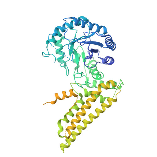
9QEN, 9QEP - PubMed Abstract:
Nucleocytoplasmic protein O-GlcNAcylation is a dynamic modification catalysed by O-GlcNAc transferase (OGT) and reversed by O-GlcNAc hydrolase (OGA), whose activities are regulated through largely unknown O-GlcNAc-dependent feedback mechanisms. OGA is a homodimeric, multi-domain enzyme containing a catalytic core and a pseudo-histone acetyltransferase (pHAT) domain. While a catalytic structure has been reported, the structure and function of the pHAT domain remain elusive. Here, we report a crystal structure of the Trichoplax adhaerens pHAT domain and cryo-EM data of the multi-domain T. adhaerens and human OGAs, complemented by biophysical analyses. Here, we show that the eukaryotic OGA pHAT domain forms catalytically incompetent, symmetric homodimers, projecting a partially conserved putative peptide-binding site. In solution, OGA exist as flexible multi-domain dimers, but catalytic core-pHAT linker interactions restrict pHAT positional range. In human OGA, pHAT movements remodel the active site environment through conformational changes in a flexible arm region. These findings reveal allosteric mechanisms through which the pHAT domain contributes to O-GlcNAc homeostasis.
- Section for Neurobiology and DANDRITE, Department of Molecular Biology and Genetics, Aarhus University, Aarhus, Denmark.
Organizational Affiliation: