Enhancing Solubility in VHL-Based PROTACs: Optimized USP7 Degraders for Improved Developability.
Wittenburg, S., Zuleeg, M.R., Peter, K., Lemnitzer, P., Voget, R., Bricelj, A., Gobec, M., Dierlamm, N., Braun, M.B., Geiger, T.M., Heim, C., Stakemeier, A., Wagner, K.G., Nowak, R.P., Hartmann, M.D., Sosic, I., Gutschow, M., Kronke, J., Steinebach, C.(2025) J Med Chem 68: 15711-15737
- PubMed: 40673806 Search on PubMed
- DOI: https://doi.org/10.1021/acs.jmedchem.5c00718
- Primary Citation Related Structures:
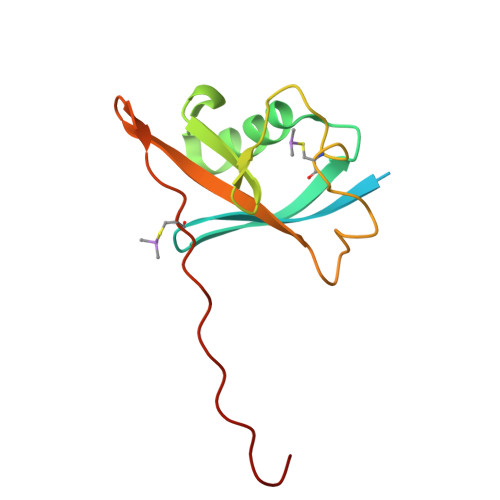
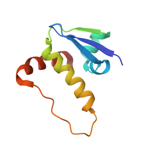
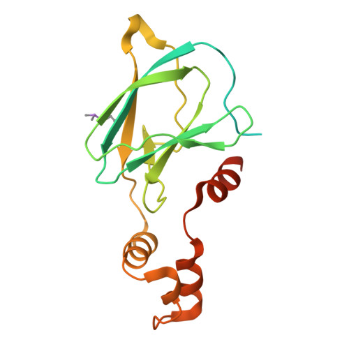
9QE4, 9QE5 - PubMed Abstract:
Limited aqueous solubility, high total polar surface area (TPSA), and high hydrogen-bond donor (HBD) counts have hampered the clinical development of VHL-based proteolysis-targeting chimeras (PROTACs). This study explores strategies to enhance the physicochemical properties of VHL-recruiting USP7 degraders. By adjusting lipophilicity, HBD count, and TPSA, we created degraders with improved solubility while maintaining their USP7 degradation capability. Structural modifications at the VHL ligand included a constrained six-membered ring in the peptidic scaffold and the addition of solubilizing groups. These changes enhanced aqueous solubility without compromising degradation performance. A key example is PROTAC 40 , modified with a dibasic piperazine, which exhibits a 170-fold increase in solubility over its predecessor while retaining strong target selectivity. The findings demonstrate that rational scaffold design can yield solubility-enhanced VHL-based PROTACs with broad potential for drug development. This methodology may also be applicable to other E3 ligases, supporting the development of degraders suitable for in vivo use.
- Pharmaceutical Institute, Department of Pharmaceutical & Medicinal Chemistry, University of Bonn, An der Immenburg 4, DE-53121 Bonn, Germany.
Organizational Affiliation: