Escherichia coli Triheme Enzyme YhjA: Structure and Reactivity.
Hewitt, P., Seidel, J., Wust, A., Smith, M., Maiocco, S.J., Shternberg, S., Hoffmann, M., Spatzal, T., Gerhardt, S., Einsle, O., Elliott, S.J.(2025) Biochemistry 64: 3322-3332
- PubMed: 40669070 Search on PubMed
- DOI: https://doi.org/10.1021/acs.biochem.5c00202
- Primary Citation Related Structures:
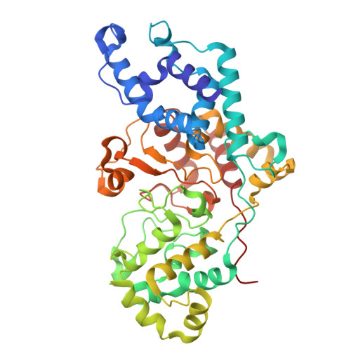
9QDT - PubMed Abstract:
It has been recently realized that some Gram-negative organisms such as Escherichia coli produce a multiheme cytochrome c to serve as a quinol peroxidase that couples electrons from the quinol pool directly to H 2 O 2 . The E. coli version of this enzyme, termed YhjA, has been predicted to be a member of the bacterial cytochrome c peroxidase (bCCP) superfamily, where a novel N-terminal single-heme binding domain is fused to the canonical bCCP diheme domain found widely in Gram-negative bacteria. Here, we present an X-ray crystal structure of YhjA, revealing the triheme architecture that nature has employed to couple the quinol pool to the reduction of H 2 O 2 . We also show kinetic, spectroscopic, and electrochemical data that detail the differences between the three hemes that are observed in the structure, where two of the heme irons are both six-coordinate, ligated by Met and His residues, and the third peroxidatic heme is found to be five-coordinate. Electrocatalytic voltammetry of YhjA illustrates how the high-potential hemes serve as relays to the peroxidatic active site. Together, these data suggest a model of the catalytic chemistry of YhjA, illustrating how this member of the bCCP family may react with substrates and engage in multielectron redox reactions.
- Department of Chemistry, Boston University, Boston, Massachusetts 02215, United States.
Organizational Affiliation: