9QB7 | pdb_00009qb7

Structure of rat Phospholipase C gamma 1 mutant S345F


Experimental Data Snapshot

  • Method: X-RAY DIFFRACTION
  • Resolution: 2.56 Å
  • R-Value Free: 
    0.233 (Depositor), 0.232 (DCC) 
  • R-Value Work: 
    0.188 (Depositor), 0.187 (DCC) 
  • R-Value Observed: 
    0.190 (Depositor) 

Starting Model: experimental
View more details

wwPDB Validation   3D Report Full Report


This is version 1.1 of the entry. See complete history


Literature

Characterisation of an allosteric site in PLC gamma enzymes and implications for development of their specific inhibitors.

Bunney, T.D.Nyvall, H.G.Macrae, C.Lalovic, D.Gregory, A.Le Huray, K.I.P.Harvey, N.Pinotsis, N.Kalli, A.C.Waudby, C.A.Burke, J.E.Katan, M.

(2025) Biochem J 482: 1545-1564

  • DOI: https://doi.org/10.1042/BCJ20253358
  • Primary Citation of Related Structures:  
    9QB7

  • PubMed Abstract: 

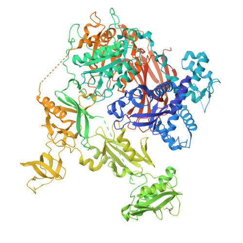
    Phospholipase C gamma (PLCγ) enzymes are key components of intracellular signal transduction processes and are involved in disease development, including immune dysregulation, specific cancer types and neurodegeneration. Although recognised as important targets for intervention, validated pharmacological tools are lacking. Here, we demonstrate that inhibitory nucleotides bind directly to an allosteric site at the interface between the PLC-core and regulatory-array unique for PLCγ, underlying their specificity for the PLCγ family. This binding site overlaps with the PLCγ autoinhibitory interface, suggesting that the inhibitory impact of nucleotides involves stabilisation of autoinhibition. We have also analysed disease-linked variants of PLCγ1 and PLCγ2 to show that multiple mechanisms could underpin their gain-of-function phenotype. While the sensitivity of these variants to physiological nucleotide inhibition is reduced, we identified artificial nucleotide compounds that can inhibit such variants not only in vitro but also in cell-based assays. Therefore, our findings suggest a route for development of isozyme specific PLCγ inhibitors allowing further studies of their roles in health and disease.


  • Organizational Affiliation
    • Institute of Structural and Molecular Biology, Division of Biosciences, University College London, London, WC1E 6BT, UK.

Macromolecules
Find similar proteins by:  (by identity cutoff)  |  3D Structure
Entity ID: 1
MoleculeChains Sequence LengthOrganismDetailsImage
1-phosphatidylinositol 4,5-bisphosphate phosphodiesterase gamma-11,268Rattus norvegicusMutation(s): 1 
Gene Names: Plcg1
EC: 3.1.4.11
UniProt
Find proteins for P10686 (Rattus norvegicus)
Explore P10686 
Go to UniProtKB:  P10686
Entity Groups  
Sequence Clusters30% Identity50% Identity70% Identity90% Identity95% Identity100% Identity
UniProt GroupP10686
Sequence Annotations
Expand
  • Reference Sequence
Small Molecules
Ligands 1 Unique
IDChains Name / Formula / InChI Key2D Diagram3D Interactions
SO4
Query on SO4

Download Ideal Coordinates CCD File 
B [auth A]SULFATE ION
O4 S
QAOWNCQODCNURD-UHFFFAOYSA-L
Experimental Data & Validation

Experimental Data

  • Method: X-RAY DIFFRACTION
  • Resolution: 2.56 Å
  • R-Value Free:  0.233 (Depositor), 0.232 (DCC) 
  • R-Value Work:  0.188 (Depositor), 0.187 (DCC) 
  • R-Value Observed: 0.190 (Depositor) 
Space Group: P 21 21 21
Unit Cell:
Length ( Å )Angle ( ˚ )
a = 72.442α = 90
b = 82.391β = 90
c = 230.676γ = 90
Software Package:
Software NamePurpose
PHENIXrefinement
autoPROCdata reduction
autoPROCdata scaling

Structure Validation

View Full Validation Report


Entry History & Funding Information

Deposition Data


Funding OrganizationLocationGrant Number
Not funded--

Revision History  (Full details and data files)

  • Version 1.0: 2025-10-29
    Type: Initial release
  • Version 1.1: 2025-12-24
    Changes: Database references