9Q3J | pdb_00009q3j

Structures of LarA-like nickel-pincer nucleotide cofactor-utilizing enzyme with a single catalytic histidine residue from Blautia wexlerae


Experimental Data Snapshot

  • Method: ELECTRON MICROSCOPY
  • Resolution: 3.15 Å
  • Aggregation State: PARTICLE 
  • Reconstruction Method: SINGLE PARTICLE 

wwPDB Validation   3D Report Full Report


This is version 1.0 of the entry. See complete history


Literature

Structures of two LarA-like nickel-pincer nucleotide cofactor-utilizing enzymes with a single catalytic histidine residue.

Gatreddi, S.Subramanian, S.Sui, D.Wang, T.Urdiain-Arraiza, J.Desguin, B.Hausinger, R.P.Parent, K.N.Hu, J.

(2025) bioRxiv 

  • DOI: https://doi.org/10.1101/2025.08.19.671153
  • Primary Citation of Related Structures:  
    9Q3J, 9Q3K

  • PubMed Abstract: 

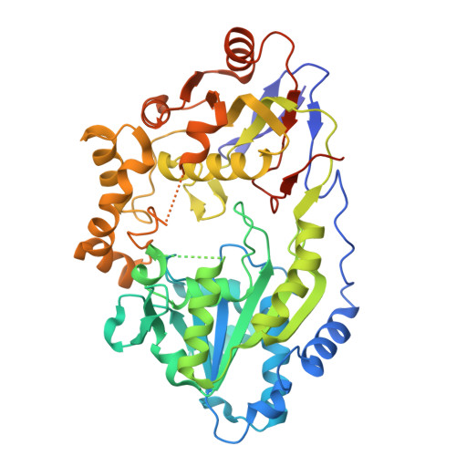
    The nickel pincer nucleotide (NPN) cofactor catalyzes the racemization/epimerization of α-hydroxy acids in enzymes of the LarA family. The established proton-coupled hydride transfer mechanism requires two catalytic histidine residues that alternately act as general acids and general bases. Notably, however, a fraction of LarA homologs (LarAHs) lack one of the active site histidine residues, replacing it with an asparaginyl side chain that cannot participate in acid/base catalysis. Here, we investigated two such LarAHs and solved their cryo-electron microscopic structures with and without loaded NPN cofactor, respectively. The structures revealed a consistent octameric assembly that is unprecedented in the LarA family and unveiled a new set of active site residues that likely recognize and process substrates differently from those of the well-studied LarAHs. Genomic context analysis suggested their potential involvement in carbohydrate metabolism. Together, these findings lay the groundwork for expanding the breadth of reactions and the range of mechanisms of LarA enzymes.


  • Organizational Affiliation
    • Department of Microbiology, Genetics, and Immunology, Michigan State University, MI, USA.

Macromolecules
Find similar proteins by:  (by identity cutoff)  |  3D Structure
Entity ID: 1
MoleculeChains Sequence LengthOrganismDetailsImage
DUF2088 domain-containing protein
A, B, C, D, E
A, B, C, D, E, F, G, H
505Blautia wexleraeMutation(s): 0 
Gene Names: ERS852523_02626GT728_05510
UniProt
Find proteins for A0A174QNZ5 (Blautia wexlerae)
Explore A0A174QNZ5 
Go to UniProtKB:  A0A174QNZ5
Entity Groups  
Sequence Clusters30% Identity50% Identity70% Identity90% Identity95% Identity100% Identity
UniProt GroupA0A174QNZ5
Sequence Annotations
Expand
  • Reference Sequence
Experimental Data & Validation

Experimental Data

  • Method: ELECTRON MICROSCOPY
  • Resolution: 3.15 Å
  • Aggregation State: PARTICLE 
  • Reconstruction Method: SINGLE PARTICLE 
EM Software:
TaskSoftware PackageVersion
MODEL REFINEMENTPHENIX1.20.1_4487:
RECONSTRUCTIONcryoSPARC

Structure Validation

View Full Validation Report


Entry History & Funding Information

Deposition Data


Funding OrganizationLocationGrant Number
National Institutes of Health/National Institute of General Medical Sciences (NIH/NIGMS)United StatesGM128959
National Institutes of Health/National Institute of General Medical Sciences (NIH/NIGMS)United StatesGM140931
National Institutes of Health/National Institute of General Medical Sciences (NIH/NIGMS)United StatesGM140803

Revision History  (Full details and data files)

  • Version 1.0: 2025-09-17
    Type: Initial release