9PRU | pdb_00009pru

Complex of the 3G8 Fab bound to Fc gamma receptor 3a / CD16a F158 allotype


Experimental Data Snapshot

  • Method: X-RAY DIFFRACTION
  • Resolution: 1.90 Å
  • R-Value Free: 
    0.280 (Depositor), 0.277 (DCC) 
  • R-Value Work: 
    0.230 (Depositor), 0.231 (DCC) 
  • R-Value Observed: 
    0.230 (Depositor) 

Starting Model: experimental
View more details

wwPDB Validation   3D Report Full Report


Ligand Structure Quality Assessment 


This is version 1.0 of the entry. See complete history


Literature

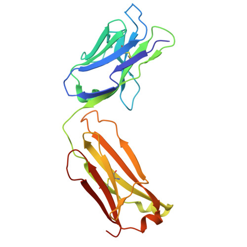
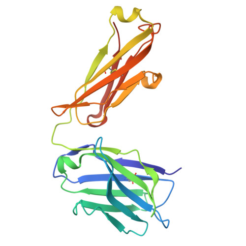
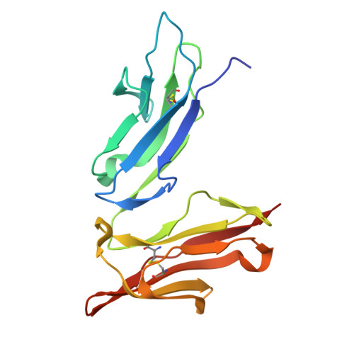
The impact of N-glycan conformational entropy on the binding affinity of Fc gamma receptor IIIa/CD16a.

Kremer, P.G.Tolbert, W.D.Gazaway, E.Hernandez, B.G.Korzeniowski, M.K.Dyba, Z.A.Grelsson, T.Grant, O.C.Lanzilotta, W.N.Pazgier, M.Woods, R.J.Barb, A.W.

(2025) Structure 

  • DOI: https://doi.org/10.1016/j.str.2025.11.015
  • Primary Citation of Related Structures:  
    7URU, 9PRU

  • PubMed Abstract: 

    The affinity of Fc γ receptor IIIa (FcγRIIIa) binding to immunoglobulin G1 (IgG1) correlates with patient responses for antibody-based therapeutics. Among multiple factors affecting affinity, a mechanism defining how the composition of the FcγRIIIa N162 glycan regulates affinity remains undefined. Here, we evaluate the binding modes of two competitive FcγRIIIa ligands. IgG1 Fc binding is sensitive to N162 glycan composition, unlike the antigen-binding fragment (Fab) of the FcγRIII-specific antibody 3G8. Both ligands bound to overlapping surfaces, utilizing different angles of attack such that the IgG1 Fc but not 3G8 Fab limited the space available to the FcγRIII N162 N-glycan. FcγRIII binding to IgG1 Fc generated a 2.1 kcal/mol penalty from a loss of N162 glycan conformational entropy, greater than the 0.3 kcal/mol penalty for 3G8 and consistent with binding measurements. Thus, the conformational entropy of the FcγRIIIa N162-glycan is the predominant force modulating differential binding affinity compared to 3G8 Fab binding for endogenous FcγRIIIa glycoforms.


  • Organizational Affiliation
    • Department of Biochemistry and Molecular Biology, University of Georgia, Athens, GA, USA.

Macromolecules
Find similar proteins by:  (by identity cutoff)  |  3D Structure
Entity ID: 1
MoleculeChains Sequence LengthOrganismDetailsImage
3G8 Fab light chain
A, C, E, G
218Mus musculusMutation(s): 0 
Entity Groups  
Sequence Clusters30% Identity50% Identity70% Identity90% Identity95% Identity100% Identity
Sequence Annotations
Expand
  • Reference Sequence
Find similar proteins by:  (by identity cutoff)  |  3D Structure
Entity ID: 2
MoleculeChains Sequence LengthOrganismDetailsImage
3G8 Fab Heavy Chain
B, D, F, H
221Mus musculusMutation(s): 0 
Entity Groups  
Sequence Clusters30% Identity50% Identity70% Identity90% Identity95% Identity100% Identity
Sequence Annotations
Expand
  • Reference Sequence
Find similar proteins by:  (by identity cutoff)  |  3D Structure
Entity ID: 3
MoleculeChains Sequence LengthOrganismDetailsImage
Low affinity immunoglobulin gamma Fc region receptor III-AI [auth W],
J [auth X],
K [auth Y],
L [auth Z]
175Homo sapiensMutation(s): 3 
Gene Names: FCGR3ACD16AFCG3FCGR3IGFR3
UniProt & NIH Common Fund Data Resources
Find proteins for P08637 (Homo sapiens)
Explore P08637 
Go to UniProtKB:  P08637
PHAROS:  P08637
GTEx:  ENSG00000203747 
Entity Groups  
Sequence Clusters30% Identity50% Identity70% Identity90% Identity95% Identity100% Identity
UniProt GroupP08637
Glycosylation
Glycosylation Sites: 2Go to GlyGen: P08637-1
Sequence Annotations
Expand
  • Reference Sequence
Oligosaccharides

Help

Entity ID: 4
MoleculeChains Length2D Diagram Glycosylation3D Interactions
beta-D-mannopyranose-(1-4)-2-acetamido-2-deoxy-beta-D-glucopyranose-(1-4)-2-acetamido-2-deoxy-beta-D-glucopyranoseM [auth I],
N [auth J],
P [auth L]
3N-Glycosylation
Entity ID: 5
MoleculeChains Length2D Diagram Glycosylation3D Interactions
2-acetamido-2-deoxy-beta-D-glucopyranose-(1-4)-2-acetamido-2-deoxy-beta-D-glucopyranoseO [auth K]2N-Glycosylation
Small Molecules
Ligands 4 Unique
IDChains Name / Formula / InChI Key2D Diagram3D Interactions
NAG (Subject of Investigation/LOI)
Query on NAG

Download Ideal Coordinates CCD File 
AA [auth Z],
X [auth W],
Y [auth X],
Z [auth Y]
2-acetamido-2-deoxy-beta-D-glucopyranose
C8 H15 N O6
OVRNDRQMDRJTHS-FMDGEEDCSA-N
GOL
Query on GOL

Download Ideal Coordinates CCD File 
Q [auth A],
R [auth C],
T [auth E]
GLYCEROL
C3 H8 O3
PEDCQBHIVMGVHV-UHFFFAOYSA-N
CL
Query on CL

Download Ideal Coordinates CCD File 
V [auth G]CHLORIDE ION
Cl
VEXZGXHMUGYJMC-UHFFFAOYSA-M
NA
Query on NA

Download Ideal Coordinates CCD File 
S [auth E],
U [auth G],
W [auth H]
SODIUM ION
Na
FKNQFGJONOIPTF-UHFFFAOYSA-N
Experimental Data & Validation

Experimental Data

  • Method: X-RAY DIFFRACTION
  • Resolution: 1.90 Å
  • R-Value Free:  0.280 (Depositor), 0.277 (DCC) 
  • R-Value Work:  0.230 (Depositor), 0.231 (DCC) 
  • R-Value Observed: 0.230 (Depositor) 
Space Group: P 21 21 21
Unit Cell:
Length ( Å )Angle ( ˚ )
a = 130.062α = 90
b = 130.594β = 90
c = 170.511γ = 90
Software Package:
Software NamePurpose
PHENIXrefinement
HKL-2000data reduction
HKL-2000data scaling
PHENIXphasing

Structure Validation

View Full Validation Report


Ligand Structure Quality Assessment 


Entry History & Funding Information

Deposition Data


Funding OrganizationLocationGrant Number
National Institutes of Health/National Institute Of Allergy and Infectious Diseases (NIH/NIAID)United StatesAI148114
National Institutes of Health/National Institute Of Allergy and Infectious Diseases (NIH/NIAID)United StatesAI187028

Revision History  (Full details and data files)

  • Version 1.0: 2025-12-31
    Type: Initial release