Structure of a transient protein-folding intermediate by pressure-jump NMR spectroscopy.
Masoumzadeh, E., Courtney, J.M., Charlier, C., Ying, J., Anfinrud, P., Bax, A.(2025) Proc Natl Acad Sci U S A 122: e2519493122-e2519493122
- PubMed: 41060762 Search on PubMed
- DOI: https://doi.org/10.1073/pnas.2519493122
- Primary Citation Related Structures:
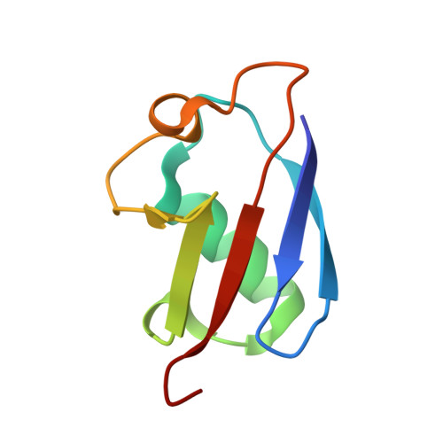
9PL1 - PubMed Abstract:
Protein folding, as commonly portrayed, involves exploration of a rough, high-dimensional landscape, ending with a final descent into a low-energy folded state. During that journey, the protein may visit shallow basins corresponding to metastable structures, potentially of biological importance. Structural characterization of transiently populated metastable states is challenging due to their low population, which limits traditional NMR, and also makes crystallization for X-ray diffraction difficult without stabilizing mutations, covalent modifications, or the addition of antibodies. Here, we report the structural characterization of the on-pathway folding intermediate of a pressure-sensitized ubiquitin mutant. The obtained non-native β-sheet registry was previously shown to be necessary in the PINK1 mitophagy pathway. We used fast pressure jumps to repeatedly initiate folding and advanced NMR measurements to probe the evolving ensemble of protein conformations. The results reported here demonstrate that the non-native β-sheet hydrogen bond registry can act as a metastable trap during protein folding. This work provides a template for future investigation of metastable conformations and protein folding with rich structural detail.
- Laboratory of Chemical Physics, National Institute of Diabetes and Digestive and Kidney Diseases, NIH, Bethesda, MD 20892-0520.
Organizational Affiliation: