9PIA | pdb_00009pia

Human glutaminase C mutant S482C


Experimental Data Snapshot

  • Method: X-RAY DIFFRACTION
  • Resolution: 2.99 Å
  • R-Value Free: 
    0.209 (Depositor), 0.211 (DCC) 
  • R-Value Work: 
    0.188 (Depositor), 0.188 (DCC) 
  • R-Value Observed: 
    0.188 (Depositor) 

Starting Model: experimental
View more details

wwPDB Validation 3D Report Full Report

Validation slider image for 9PIA

This is version 1.1 of the entry. See complete history

Literature

Structure and enzymology of glutaminase S482C and H461L variants associated with excess brain glutamate and neurological disease.

Crane, C.S.McIssac, T.K.Milano, S.K.Cerione, R.A.Ulrich, S.M.

(2026) J Biological Chem : 113091-113091

  • DOI: https://doi.org/10.1016/j.jbc.2026.113091
  • Primary Citation Related Structures: 
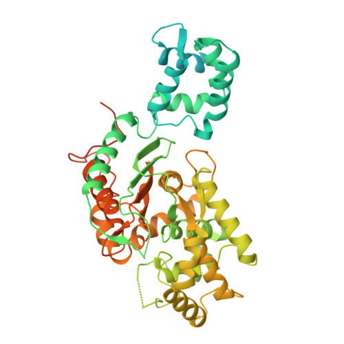
    9PIA

  • PubMed Abstract: 

    Glutaminase (GLS) catalyzes the hydrolysis of glutamine to produce glutamate, the brain's principal excitatory neurotransmitter. Two de novo gain-of-function mutations in GLS, S482C and H461L, were recently identified in patients with developmental delay, epilepsy, and infantile cataract. These patients exhibited high glutamate and low glutamine concentrations in the brain, suggesting that the GLS variants have abnormal enzymology. Here, we examined the enzymatic properties of the mutant enzymes and found that they no longer require the anionic activator phosphate to stimulate enzymatic activity or induce filament formation. The mutant enzymes also exhibit a total (S482C) or partial (H461L) loss of glutamate product inhibition, lifting this restriction on glutamate accumulation. Structural analysis of the S482C variant shows the mutation shifts the key catalytic residue Y466 into its catalytically active configuration and disrupts a key hydrogen bond between Y466 and the glutamate product, explaining how the S482C variant has enzymatic activity in the absence of phosphate and is insensitive to glutamate product inhibition. These results shed new light on the mechanism of phosphate activation and glutamate product inhibition of GLS and show that loss of these enzymatic properties disrupts glutamate homeostasis in the brain and causes neurological disease.


  • Organizational Affiliation
    • Department of Chemistry and Chemical Biology, Cornell University, Ithaca, NY 14853.

Macromolecule Content 

  • Total Structure Weight: 262.23 kDa 
  • Atom Count: 12,492 
  • Modeled Residue Count: 1,592 
  • Deposited Residue Count: 2,392 
  • Unique protein chains: 1

Macromolecules

Find similar proteins by:|  3D Structure
Entity ID: 1
MoleculeChains  Sequence LengthOrganismDetailsImage
Isoform 3 of Glutaminase kidney isoform, mitochondrial
A, B, C, D
598Homo sapiensMutation(s): 1 
Gene Names: GLSGLS1KIAA0838
EC: 3.5.1.2
UniProt & NIH Common Fund Data Resources
Find proteins for O94925 (Homo sapiens)
Explore O94925 
Go to UniProtKB:  O94925
PHAROS:  O94925
GTEx:  ENSG00000115419 
Entity Groups
Sequence Clusters30% Identity50% Identity70% Identity90% Identity95% Identity100% Identity
UniProt GroupO94925
Sequence Annotations
Expand
Reference Sequence

Experimental Data & Validation

Experimental Data

  • Method: X-RAY DIFFRACTION
  • Resolution: 2.99 Å
  • R-Value Free:  0.209 (Depositor), 0.211 (DCC) 
  • R-Value Work:  0.188 (Depositor), 0.188 (DCC) 
  • R-Value Observed: 0.188 (Depositor) 
Space Group: P 1 21 1
Unit Cell:
Length ( Å )Angle ( ˚ )
a = 50.851α = 90
b = 138.209β = 93.58
c = 177.122γ = 90
Software Package:
Software NamePurpose
PHENIXrefinement
HKL-2000data scaling
HKL-2000data reduction
PDB_EXTRACTdata extraction
PHASERphasing

Structure Validation

View Full Validation Report


Entry History 

& Funding Information

Deposition Data


Funding OrganizationLocationGrant Number
National Institutes of Health/National Cancer Institute (NIH/NCI)United States--

Revision History  (Full details and data files)

  • Version 1.0: 2025-10-01
    Type: Initial release
  • Version 1.1: 2026-05-13
    Changes: Database references