9OMX | pdb_00009omx

Crystal structure of E. coli ApaH in complex with Up4U


Experimental Data Snapshot

  • Method: X-RAY DIFFRACTION
  • Resolution: 1.54 Å
  • R-Value Free: 
    0.196 (Depositor), 0.196 (DCC) 
  • R-Value Work: 
    0.177 (Depositor), 0.178 (DCC) 
  • R-Value Observed: 
    0.178 (Depositor) 

Starting Model: experimental
View more details

wwPDB Validation 3D Report Full Report

Validation slider image for 9OMX

Ligand Structure Quality Assessment 


This is version 1.0 of the entry. See complete history

Literature

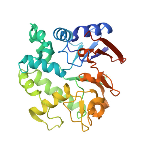
ApaH decaps Np 4 N-capped RNAs in two alternative orientations.

Nuthanakanti, A.Korn, M.Levenson-Palmer, R.Wu, Y.Babu, N.R.Huang, X.Banh, R.S.Belasco, J.G.Serganov, A.

(2025) Nat Chem Biol 

  • DOI: https://doi.org/10.1038/s41589-025-01991-4
  • Primary Citation Related Structures: 
    9OJD, 9OJP, 9OJQ, 9OJW, 9OJX, 9OK1, 9OK2, 9OLN, 9OLY, 9OLZ, 9OM9, 9OMC, 9OMU, 9OMW, 9OMX, 9ON0, 9ON7, 9OND, 9ONG, 9OON, 9OOY, 9OP2, 9OPG, 9OPH, 9OQ9, 9OQB

  • PubMed Abstract: 

    Enigmatic dinucleoside tetraphosphates, known as 'alarmones' (Np 4 Ns), have recently been shown to function in bacteria as precursors to Np 4 caps on transcripts, likely influencing RNA longevity and cellular adaptation to stress. In proteobacteria, ApaH is the predominant enzyme that hydrolyzes Np 4 Ns and decaps Np 4 -capped RNAs to initiate their 5'-end-dependent degradation. Here we conducted a biochemical and structural study to uncover the catalytic mechanism of Escherichia coli ApaH, a prototypic symmetric Np 4 N hydrolase, on various Np 4 Ns and Np 4 -capped RNAs. We found that the enzyme uses a unique combination of nonspecific and semispecific substrate recognition, enabling substrates to bind in two orientations with a slight orientational preference. Despite such exceptional recognition properties, ApaH efficiently decaps various Np 4 -capped mRNAs and sRNAs, thereby impacting their lifetimes. Our findings highlight the need to determine substrate orientation preferences before designing substrate-mimicking drugs, as enzymes may escape activity modulation with one of the alternative substrate orientations.


  • Organizational Affiliation
    • Department of Biochemistry and Molecular Pharmacology, New York University Grossman School of Medicine, New York City, NY, USA.

Macromolecule Content 

  • Total Structure Weight: 66.89 kDa 
  • Atom Count: 5,144 
  • Modeled Residue Count: 538 
  • Deposited Residue Count: 572 
  • Unique protein chains: 1

Macromolecules

Find similar proteins by:|  3D Structure
Entity ID: 1
MoleculeChains  Sequence LengthOrganismDetailsImage
Bis(5'-nucleosyl)-tetraphosphatase [symmetrical]
A, B
286Escherichia coli K-12Mutation(s): 0 
Gene Names: apaHb0049JW0048
EC: 3.6.1.41
UniProt
Find proteins for P05637 (Escherichia coli (strain K12))
Explore P05637 
Go to UniProtKB:  P05637
Entity Groups
Sequence Clusters30% Identity50% Identity70% Identity90% Identity95% Identity100% Identity
UniProt GroupP05637
Sequence Annotations
Expand
Reference Sequence

Small Molecules

Ligands 5 Unique
IDChains Name / Formula / InChI Key2D Diagram3D Interactions
A1CC9
(Subject of Investigation/LOI)

Query on A1CC9


Download:Ideal Coordinates CCD File
C [auth A],
K [auth B]
[[(2~{R},3~{S},4~{R},5~{R})-5-[2,4-bis(oxidanylidene)pyrimidin-1-yl]-3,4-bis(oxidanyl)oxolan-2-yl]methoxy-oxidanyl-phosphoryl] [[[(3~{S},4~{S},5~{R})-5-[2,4-bis(oxidanylidene)pyrimidin-1-yl]-3,4-bis(oxidanyl)oxolan-2-yl]methoxy-oxidanyl-phosphoryl]oxy-oxidanyl-phosphoryl] hydrogen phosphate
C18 H26 N4 O23 P4
NMLMACJWHPHKGR-JILDPCHGSA-N
EPE

Query on EPE


Download:Ideal Coordinates CCD File
J [auth B]4-(2-HYDROXYETHYL)-1-PIPERAZINE ETHANESULFONIC ACID
C8 H18 N2 O4 S
JKMHFZQWWAIEOD-UHFFFAOYSA-N
SO4

Query on SO4


Download:Ideal Coordinates CCD File
G [auth A],
H [auth A],
I [auth A],
O [auth B],
P [auth B]
SULFATE ION
O4 S
QAOWNCQODCNURD-UHFFFAOYSA-L
MN

Query on MN


Download:Ideal Coordinates CCD File
D [auth A],
F [auth A],
L [auth B],
N [auth B]
MANGANESE (II) ION
Mn
WAEMQWOKJMHJLA-UHFFFAOYSA-N
MG

Query on MG


Download:Ideal Coordinates CCD File
E [auth A],
M [auth B]
MAGNESIUM ION
Mg
JLVVSXFLKOJNIY-UHFFFAOYSA-N

Experimental Data & Validation

Experimental Data

  • Method: X-RAY DIFFRACTION
  • Resolution: 1.54 Å
  • R-Value Free:  0.196 (Depositor), 0.196 (DCC) 
  • R-Value Work:  0.177 (Depositor), 0.178 (DCC) 
  • R-Value Observed: 0.178 (Depositor) 
Space Group: C 1 2 1
Unit Cell:
Length ( Å )Angle ( ˚ )
a = 166.727α = 90
b = 54.964β = 129.93
c = 120.435γ = 90
Software Package:
Software NamePurpose
PHENIXrefinement
HKL-2000data scaling
HKL-2000data reduction
Cootmodel building
PHASERphasing

Structure Validation

View Full Validation Report


Ligand Structure Quality Assessment 


Entry History 

& Funding Information

Deposition Data


Funding OrganizationLocationGrant Number
National Institutes of Health/National Institute of General Medical Sciences (NIH/NIGMS)United StatesR01 GM112940

Revision History  (Full details and data files)

  • Version 1.0: 2025-09-03
    Type: Initial release