9OG0 | pdb_00009og0

Cryo-EM structure of OS9-SEL1L-HRD1 dimer


Experimental Data Snapshot

  • Method: ELECTRON MICROSCOPY
  • Resolution: 3.64 Å
  • Aggregation State: PARTICLE 
  • Reconstruction Method: SINGLE PARTICLE 

wwPDB Validation   3D Report Full Report


This is version 1.0 of the entry. See complete history


Literature

Structural basis and pathological implications of the dimeric OS9-SEL1L-HRD1 ERAD Core Complex.

Lin, L.L.Maldosevic, E.Zhou, L.E.Jomaa, A.Qi, L.

(2025) bioRxiv 

  • DOI: https://doi.org/10.1101/2025.06.13.659592
  • Primary Citation of Related Structures:  
    9OG0

  • PubMed Abstract: 

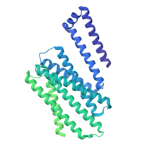
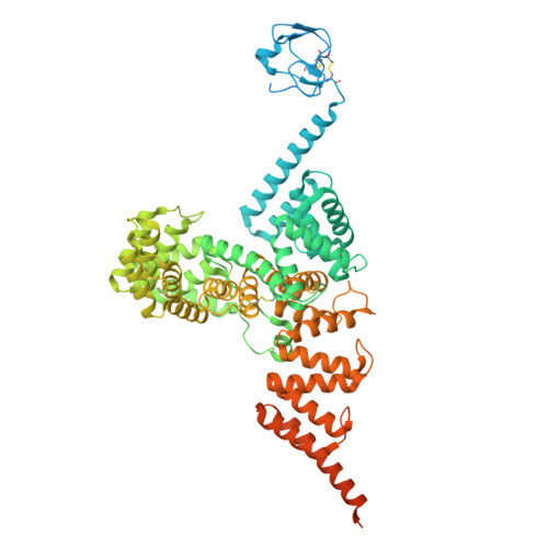
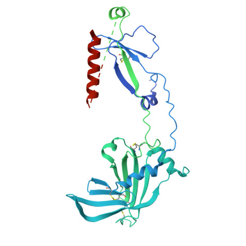
    The SEL1L-HRD1 complex represents the most conserved branch of endoplasmic reticulum (ER)-associated degradation (ERAD), a critical pathway that clears misfolded proteins to maintain ER proteostasis. However, the molecular organization and pathogenic mechanisms of mammalian ERAD have remained elusive. Here, we report the first cryo-EM structure of the core mammalian ERAD complex, comprising the ER lectin OS9, SEL1L, and the E3 ubiquitin ligase HRD1. The structure, validated by mutagenesis and crosslinking assays, reveals a dimeric assembly of the core complex in which SEL1L and OS9 form a claw-like configuration in the ER lumen that mediates substrate engagement, while HRD1 dimerizes within the membrane to facilitate substrate translocation. Pathogenic SEL1L mutations at the SEL1L-OS9 (Gly585Asp) and SEL1L-HRD1 (Ser658Pro) interfaces disrupt complex formation and impair ERAD activity. A newly identified disease-associated HRD1 variant (Ala91Asp), located in transmembrane helix 3, impairs HRD1 dimerization and substrate processing, underscoring the functional necessity of this interface and HRD1 dimerization. Finally, the structure also reveals two methionine-rich crevices flanking the HRD1 dimer, suggestive of substrate-conducting channels analogous to those in the ER membrane protein complex (EMC). These findings establish a structural framework for mammalian ERAD and elucidate how mutations destabilizing this machinery contribute to human disease. The dimeric structure of the human SEL1L-HRD1 ERAD core complex reveals key architectural and functional principles underlying the recognition and processing of misfolded proteins linked to human disease.


Macromolecules
Find similar proteins by:  (by identity cutoff)  |  3D Structure
Entity ID: 1
MoleculeChains Sequence LengthOrganismDetailsImage
E3 ubiquitin-protein ligase synoviolin
A, B
617Homo sapiensMutation(s): 0 
Gene Names: SYVN1HRD1KIAA1810
EC: 2.3.2.27
UniProt & NIH Common Fund Data Resources
Find proteins for Q86TM6 (Homo sapiens)
Explore Q86TM6 
Go to UniProtKB:  Q86TM6
PHAROS:  Q86TM6
GTEx:  ENSG00000162298 
Entity Groups  
Sequence Clusters30% Identity50% Identity70% Identity90% Identity95% Identity100% Identity
UniProt GroupQ86TM6
Sequence Annotations
Expand
  • Reference Sequence
Find similar proteins by:  (by identity cutoff)  |  3D Structure
Entity ID: 2
MoleculeChains Sequence LengthOrganismDetailsImage
Protein sel-1 homolog 1
C, D
790Mus musculusMutation(s): 0 
Gene Names: Sel1lSel1h
UniProt & NIH Common Fund Data Resources
Find proteins for Q9Z2G6 (Mus musculus)
Explore Q9Z2G6 
Go to UniProtKB:  Q9Z2G6
IMPC:  MGI:1329016
Entity Groups  
Sequence Clusters30% Identity50% Identity70% Identity90% Identity95% Identity100% Identity
UniProt GroupQ9Z2G6
Sequence Annotations
Expand
  • Reference Sequence
Find similar proteins by:  (by identity cutoff)  |  3D Structure
Entity ID: 3
MoleculeChains Sequence LengthOrganismDetailsImage
Isoform 2 of Protein OS-9
E, F
612Homo sapiensMutation(s): 0 
Gene Names: OS9
UniProt & NIH Common Fund Data Resources
Find proteins for Q13438 (Homo sapiens)
Explore Q13438 
Go to UniProtKB:  Q13438
PHAROS:  Q13438
GTEx:  ENSG00000135506 
Entity Groups  
Sequence Clusters30% Identity50% Identity70% Identity90% Identity95% Identity100% Identity
UniProt GroupQ13438
Sequence Annotations
Expand
  • Reference Sequence
Experimental Data & Validation

Experimental Data

  • Method: ELECTRON MICROSCOPY
  • Resolution: 3.64 Å
  • Aggregation State: PARTICLE 
  • Reconstruction Method: SINGLE PARTICLE 
EM Software:
TaskSoftware PackageVersion
MODEL REFINEMENTPHENIX1.20.1_4487:

Structure Validation

View Full Validation Report


Entry History & Funding Information

Deposition Data


Funding OrganizationLocationGrant Number
National Institutes of Health/National Institute of General Medical Sciences (NIH/NIGMS)United States1R35GM130292

Revision History  (Full details and data files)

  • Version 1.0: 2025-07-30
    Type: Initial release