9O6K | pdb_00009o6k

Cryo-EM structure of AMT1-AMT7-AMTP1-AMTP2 complex


Experimental Data Snapshot

  • Method: ELECTRON MICROSCOPY
  • Resolution: 3.59 Å
  • Aggregation State: PARTICLE 
  • Reconstruction Method: SINGLE PARTICLE 

wwPDB Validation   3D Report Full Report


This is version 1.1 of the entry. See complete history


Literature

Structural insight into the substrate binding of the AMT complex via an inhibitor-trapped state.

Shao, Z.Yoon, S.Lu, J.Athavale, P.Liu, Y.Song, J.

(2025) Protein Sci 34: e70265-e70265

  • DOI: https://doi.org/10.1002/pro.70265
  • Primary Citation of Related Structures:  
    9O6K

  • PubMed Abstract: 

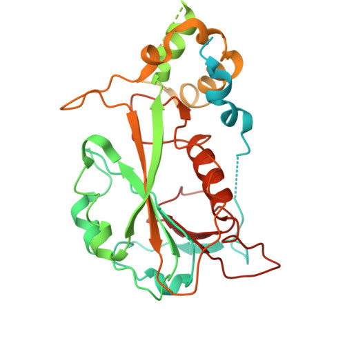
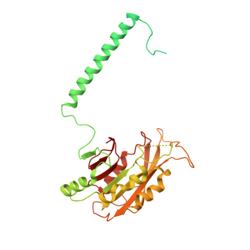
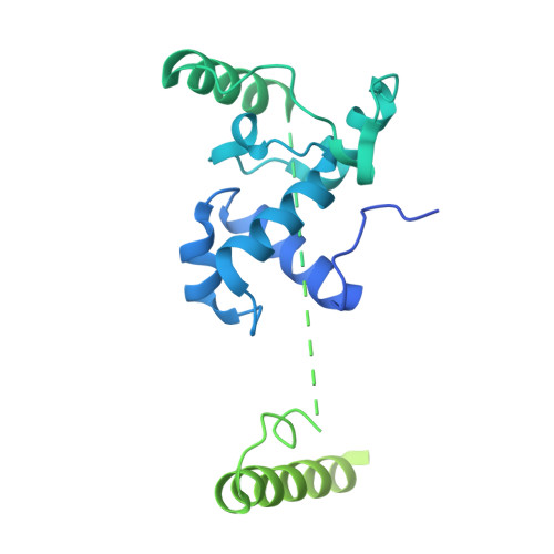
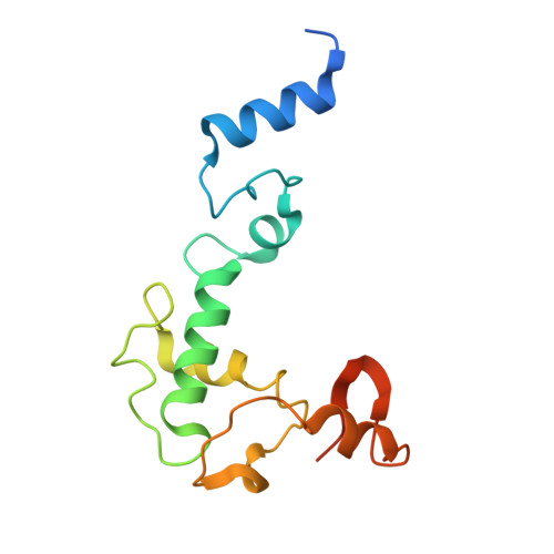
    N6-adenine (6mA) DNA methylation plays an important role in gene regulation and genome stability. The 6mA methylation in Tetrahymena thermophila is mainly mediated by the AMT complex, comprised of the AMT1, AMT7, AMTP1, and AMTP2 subunits. To date, how this complex assembles on the DNA substrate remains elusive. Here we report the structure of the AMT complex bound to the OCR protein from bacteriophage T7, mimicking the AMT-DNA encounter complex. The AMT1-AMT7 heterodimer approaches OCR from one side, while the AMTP1 N-terminal domain, assuming a homeodomain fold, binds to OCR from the other side, resulting in a saddle-shaped architecture reminiscent of what was observed for prokaryotic 6mA writers. Mutation of the AMT1, AMT7, and AMTP1 residues on the OCR-contact points led to impaired DNA methylation activity to various extents, supporting a role for these residues in DNA binding. Furthermore, structural comparison of the AMT1-AMT7 subunits with the evolutionarily related METTL3-METTL14 and AMT1-AMT6 complexes reveals sequence conservation and divergence in the region corresponding to the OCR-binding site, shedding light on the substrate binding of the latter two complexes. Together, this study supports a model in which the AMT complex undergoes a substrate binding-induced open-to-closed conformational transition, with implications in its substrate binding and processive 6mA methylation.


  • Organizational Affiliation
    • Department of Biochemistry, University of California, Riverside, California, USA.

Macromolecules
Find similar proteins by:  (by identity cutoff)  |  3D Structure
Entity ID: 1
MoleculeChains Sequence LengthOrganismDetailsImage
AMT7450Tetrahymena thermophila SB210Mutation(s): 0 
Gene Names: TTHERM_00301770
UniProt
Find proteins for I7MIF9 (Tetrahymena thermophila (strain SB210))
Explore I7MIF9 
Go to UniProtKB:  I7MIF9
Entity Groups  
Sequence Clusters30% Identity50% Identity70% Identity90% Identity95% Identity100% Identity
UniProt GroupI7MIF9
Sequence Annotations
Expand
  • Reference Sequence
Find similar proteins by:  (by identity cutoff)  |  3D Structure
Entity ID: 2
MoleculeChains Sequence LengthOrganismDetailsImage
mRNA m(6)A methyltransferase372Tetrahymena thermophila SB210Mutation(s): 0 
Gene Names: TTHERM_00704040
EC: 2.1.1.348
UniProt
Find proteins for Q22GC0 (Tetrahymena thermophila (strain SB210))
Explore Q22GC0 
Go to UniProtKB:  Q22GC0
Entity Groups  
Sequence Clusters30% Identity50% Identity70% Identity90% Identity95% Identity100% Identity
UniProt GroupQ22GC0
Sequence Annotations
Expand
  • Reference Sequence
Find similar proteins by:  (by identity cutoff)  |  3D Structure
Entity ID: 3
MoleculeChains Sequence LengthOrganismDetailsImage
Myb-like domain-containing protein360Tetrahymena thermophila SB210Mutation(s): 0 
Gene Names: TTHERM_00161750
UniProt
Find proteins for Q22VV9 (Tetrahymena thermophila (strain SB210))
Explore Q22VV9 
Go to UniProtKB:  Q22VV9
Entity Groups  
Sequence Clusters30% Identity50% Identity70% Identity90% Identity95% Identity100% Identity
UniProt GroupQ22VV9
Sequence Annotations
Expand
  • Reference Sequence
Find similar proteins by:  (by identity cutoff)  |  3D Structure
Entity ID: 4
MoleculeChains Sequence LengthOrganismDetailsImage
AMTP2D [auth H]142Tetrahymena thermophila SB210Mutation(s): 0 
Gene Names: TTHERM_00439330
UniProt
Find proteins for I7M8B9 (Tetrahymena thermophila (strain SB210))
Explore I7M8B9 
Go to UniProtKB:  I7M8B9
Entity Groups  
Sequence Clusters30% Identity50% Identity70% Identity90% Identity95% Identity100% Identity
UniProt GroupI7M8B9
Sequence Annotations
Expand
  • Reference Sequence
Small Molecules
Ligands 1 Unique
IDChains Name / Formula / InChI Key2D Diagram3D Interactions
SAH (Subject of Investigation/LOI)
Query on SAH

Download Ideal Coordinates CCD File 
E [auth B]S-ADENOSYL-L-HOMOCYSTEINE
C14 H20 N6 O5 S
ZJUKTBDSGOFHSH-WFMPWKQPSA-N
Experimental Data & Validation

Experimental Data

  • Method: ELECTRON MICROSCOPY
  • Resolution: 3.59 Å
  • Aggregation State: PARTICLE 
  • Reconstruction Method: SINGLE PARTICLE 
EM Software:
TaskSoftware PackageVersion
MODEL REFINEMENTPHENIX1.21.2_5419:

Structure Validation

View Full Validation Report


Entry History & Funding Information

Deposition Data


Funding OrganizationLocationGrant Number
National Institutes of Health/National Institute of General Medical Sciences (NIH/NIGMS)United States--

Revision History  (Full details and data files)

  • Version 1.0: 2025-08-13
    Type: Initial release
  • Version 1.1: 2026-02-25
    Changes: Data collection, Database references