9O06 | pdb_00009o06

Structure of human MAIT A-F7 TCR in complex with human MR1-10-formylmethylflavin


Experimental Data Snapshot

  • Method: X-RAY DIFFRACTION
  • Resolution: 2.00 Å
  • R-Value Free: 
    0.210 (Depositor), 0.211 (DCC) 
  • R-Value Work: 
    0.173 (Depositor), 0.174 (DCC) 
  • R-Value Observed: 
    0.175 (Depositor) 

Starting Model: experimental
View more details

wwPDB Validation   3D Report Full Report


Ligand Structure Quality Assessment 


This is version 1.1 of the entry. See complete history


Literature

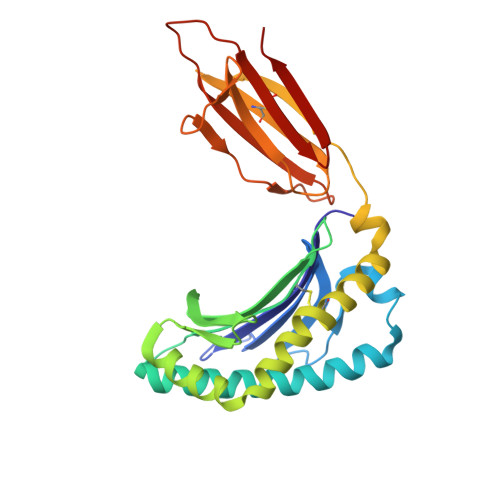
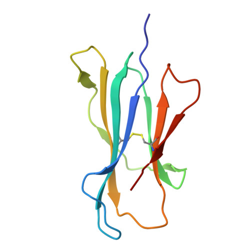
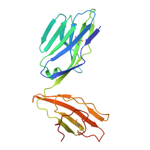
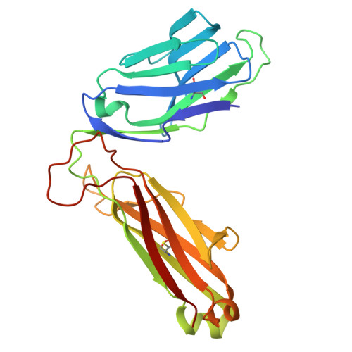
The antigen-presenting molecule MR1 binds host-generated riboflavin catabolites.

Abdelaal, M.R.Deng, J.McInerney, M.P.Ito, E.Purcell, A.W.Yamasaki, S.Villadangos, J.A.McWilliam, H.E.G.Gherardin, N.A.Rossjohn, J.Awad, W.

(2026) J Exp Medicine 223

  • DOI: https://doi.org/10.1084/jem.20250711
  • Primary Citation of Related Structures:  
    9O05, 9O06, 9O07, 9O08

  • PubMed Abstract: 

    MHC class I-related protein (MR1) presents vitamin B-based antigens (Ags) to mucosal-associated invariant T (MAIT) cells. While microbial riboflavin (RF) precursors are well-documented MR1 ligands, it is unclear whether host-generated RF catabolites influence MR1 immunity. Here, we report that RF catabolites, including 10-formylmethylflavin (FMF), lumichrome, lumiflavin, and alloxazine, bind to MR1 with moderate affinity, while RF itself binds weakly. In contrast to the MR1-upregulating microbial RF precursors, RF catabolites reduced the surface level of MR1 by inducing its retention in the endoplasmic reticulum and inhibiting exit. These RF catabolites weakly competed with vitamin B-based Ags for MR1 binding, thereby selectively inhibiting MAIT activation. The crystal structures of MR1 with RF, FMF, lumiflavin, and lumichrome show binding in the A'-pocket of MR1. Here, lumichrome formed a "flavin bond" covalent interaction with MR1-Lys43 differing from the typical Schiff base. Collectively, we identified three-ringed isoalloxazines that bind MR1 and reduce surface levels, suggesting a potential role in dampening MAIT cell immunity.


  • Organizational Affiliation
    • Infection and Immunity Program and Department of Biochemistry and Molecular Biology, Biomedicine Discovery Institute, Monash University, Clayton, Australia.

Macromolecules
Find similar proteins by:  (by identity cutoff)  |  3D Structure
Entity ID: 1
MoleculeChains Sequence LengthOrganismDetailsImage
Major histocompatibility complex class I-related gene protein
A, C
271Homo sapiensMutation(s): 1 
Gene Names: MR1
UniProt & NIH Common Fund Data Resources
Find proteins for Q95460 (Homo sapiens)
Explore Q95460 
Go to UniProtKB:  Q95460
PHAROS:  Q95460
GTEx:  ENSG00000153029 
Entity Groups  
Sequence Clusters30% Identity50% Identity70% Identity90% Identity95% Identity100% Identity
UniProt GroupQ95460
Sequence Annotations
Expand
  • Reference Sequence
Find similar proteins by:  (by identity cutoff)  |  3D Structure
Entity ID: 2
MoleculeChains Sequence LengthOrganismDetailsImage
Beta-2-microglobulin
B, F
100Homo sapiensMutation(s): 0 
Gene Names: B2MCDABP0092HDCMA22P
UniProt & NIH Common Fund Data Resources
Find proteins for P61769 (Homo sapiens)
Explore P61769 
Go to UniProtKB:  P61769
PHAROS:  P61769
GTEx:  ENSG00000166710 
Entity Groups  
Sequence Clusters30% Identity50% Identity70% Identity90% Identity95% Identity100% Identity
UniProt GroupP61769
Sequence Annotations
Expand
  • Reference Sequence
Find similar proteins by:  (by identity cutoff)  |  3D Structure
Entity ID: 3
MoleculeChains Sequence LengthOrganismDetailsImage
TCR-alpha
D, G
204Homo sapiensMutation(s): 0 
Entity Groups  
Sequence Clusters30% Identity50% Identity70% Identity90% Identity95% Identity100% Identity
Sequence Annotations
Expand
  • Reference Sequence
Find similar proteins by:  (by identity cutoff)  |  3D Structure
Entity ID: 4
MoleculeChains Sequence LengthOrganismDetailsImage
TCR-beta
E, H
246Homo sapiensMutation(s): 0 
Entity Groups  
Sequence Clusters30% Identity50% Identity70% Identity90% Identity95% Identity100% Identity
Sequence Annotations
Expand
  • Reference Sequence
Small Molecules
Ligands 3 Unique
IDChains Name / Formula / InChI Key2D Diagram3D Interactions
A1B7N (Subject of Investigation/LOI)
Query on A1B7N

Download Ideal Coordinates CCD File 
I [auth A],
L [auth C]
(7,8-dimethyl-2,4-dioxo-3,4-dihydrobenzo[g]pteridin-10(2H)-yl)acetaldehyde
C14 H12 N4 O3
AUZWNAFEXXLJDA-UHFFFAOYSA-N
GOL
Query on GOL

Download Ideal Coordinates CCD File 
J [auth A],
K [auth A],
M [auth F],
N [auth F]
GLYCEROL
C3 H8 O3
PEDCQBHIVMGVHV-UHFFFAOYSA-N
NA
Query on NA

Download Ideal Coordinates CCD File 
O [auth F],
P [auth H]
SODIUM ION
Na
FKNQFGJONOIPTF-UHFFFAOYSA-N
Experimental Data & Validation

Experimental Data

  • Method: X-RAY DIFFRACTION
  • Resolution: 2.00 Å
  • R-Value Free:  0.210 (Depositor), 0.211 (DCC) 
  • R-Value Work:  0.173 (Depositor), 0.174 (DCC) 
  • R-Value Observed: 0.175 (Depositor) 
Space Group: C 1 2 1
Unit Cell:
Length ( Å )Angle ( ˚ )
a = 217.417α = 90
b = 70.319β = 104.632
c = 143.284γ = 90
Software Package:
Software NamePurpose
PHENIXrefinement
XDSdata reduction
XDSdata scaling
PHASERphasing

Structure Validation

View Full Validation Report


Ligand Structure Quality Assessment 


Entry History & Funding Information

Deposition Data


Funding OrganizationLocationGrant Number
Australian Research Council (ARC)Australia--

Revision History  (Full details and data files)

  • Version 1.0: 2025-11-19
    Type: Initial release
  • Version 1.1: 2025-12-24
    Changes: Database references