9NZH | pdb_00009nzh

Crystal Structure of Amylin-NHO-18 binder complex


Experimental Data Snapshot

  • Method: X-RAY DIFFRACTION
  • Resolution: 2.03 Å
  • R-Value Free: 
    0.235 (Depositor), 0.235 (DCC) 
  • R-Value Work: 
    0.184 (Depositor), 0.184 (DCC) 
  • R-Value Observed: 
    0.190 (Depositor) 

Starting Model: in silico
View more details

wwPDB Validation   3D Report Full Report


This is version 1.2 of the entry. See complete history


Literature

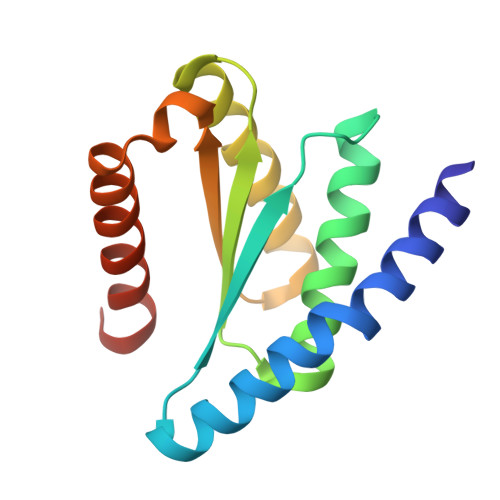
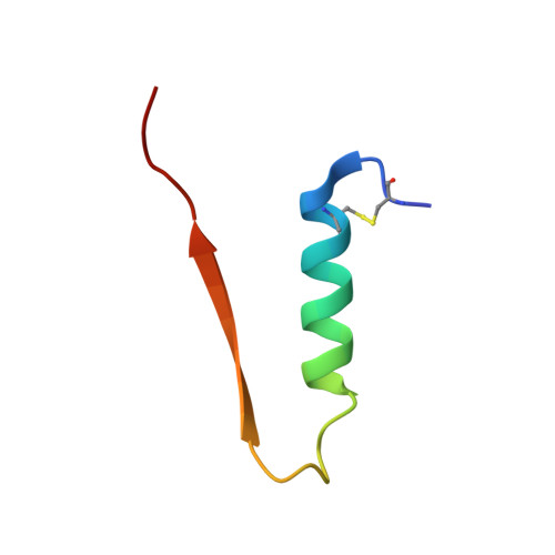
Diffusing protein binders to intrinsically disordered proteins.

Liu, C.Wu, K.Choi, H.Han, H.L.Zhang, X.Watson, J.L.Ahn, G.Zhang, J.Z.Shijo, S.Good, L.L.Fischer, C.M.Bera, A.K.Kang, A.Brackenbrough, E.Coventry, B.Hick, D.R.Qamar, S.Li, X.Decarreau, J.Gerben, S.R.Yang, W.Goreshnik, I.Vafeados, D.Wang, X.Lamb, M.Murray, A.Kenny, S.Bauer, M.S.Hoofnagle, A.N.Zhu, P.Knowles, T.P.J.Baker, D.

(2025) Nature 644: 809-817

  • DOI: https://doi.org/10.1038/s41586-025-09248-9
  • Primary Citation of Related Structures:  
    9CC5, 9CC6, 9NZH

  • PubMed Abstract: 

    Proteins that bind to intrinsically disordered proteins (IDPs) and intrinsically disordered regions (IDRs) with high affinity and specificity could be useful for therapeutic and diagnostic applications 1-4 . However, a general methodology for targeting IDPs or IDRs has yet to be developed. Here we show that starting only from the target sequence of the input, and freely sampling both target and binding protein conformations, RFdiffusion 5 can generate binders to IDPs and IDRs in a wide range of conformations. We used this approach to generate binders to the IDPs amylin, C-peptide, VP48 and BRCA1_ARATH in diverse conformations with a dissociation constant (K d ) ranging from 3 to 100 nM. For the IDRs G3BP1, common cytokine receptor γ-chain (IL-2RG) and prion protein, we diffused binders to β-strand conformations of the targets, obtaining K d between 10 and 100 nM. Fluorescence imaging experiments show that the binders bind to their respective targets in cells. The G3BP1 binder disrupts stress granule formation in cells, and the amylin binder inhibits amyloid fibril formation and dissociates existing fibres, enables targeting of both monomeric and fibrillar amylin to lysosomes, and increases the sensitivity of mass spectrometry-based amylin detection. Our approach should be useful for creating binders to flexible IDPs or IDRs spanning a wide range of intrinsic conformational preferences.


  • Organizational Affiliation
    • Department of Biochemistry, University of Washington, Seattle, WA, USA.

Macromolecules
Find similar proteins by:  (by identity cutoff)  |  3D Structure
Entity ID: 1
MoleculeChains Sequence LengthOrganismDetailsImage
Amylin-NHO-18 Binder140synthetic constructMutation(s): 0 
Entity Groups  
Sequence Clusters30% Identity50% Identity70% Identity90% Identity95% Identity100% Identity
Sequence Annotations
Expand
  • Reference Sequence
Find similar proteins by:  (by identity cutoff)  |  3D Structure
Entity ID: 2
MoleculeChains Sequence LengthOrganismDetailsImage
Islet amyloid polypeptide37Homo sapiensMutation(s): 0 
UniProt & NIH Common Fund Data Resources
Find proteins for P10997 (Homo sapiens)
Explore P10997 
Go to UniProtKB:  P10997
PHAROS:  P10997
GTEx:  ENSG00000121351 
Entity Groups  
Sequence Clusters30% Identity50% Identity70% Identity90% Identity95% Identity100% Identity
UniProt GroupP10997
Sequence Annotations
Expand
  • Reference Sequence
Experimental Data & Validation

Experimental Data

  • Method: X-RAY DIFFRACTION
  • Resolution: 2.03 Å
  • R-Value Free:  0.235 (Depositor), 0.235 (DCC) 
  • R-Value Work:  0.184 (Depositor), 0.184 (DCC) 
  • R-Value Observed: 0.190 (Depositor) 
Space Group: P 32 2 1
Unit Cell:
Length ( Å )Angle ( ˚ )
a = 59.189α = 90
b = 59.189β = 90
c = 90.946γ = 120
Software Package:
Software NamePurpose
PHENIXrefinement
XDSdata reduction
XSCALEdata scaling
PHASERphasing

Structure Validation

View Full Validation Report


Entry History & Funding Information

Deposition Data


Funding OrganizationLocationGrant Number
National Science Foundation (NSF, United States)United States--
Howard Hughes Medical Institute (HHMI)United States--

Revision History  (Full details and data files)

  • Version 1.0: 2025-05-14
    Type: Initial release
  • Version 1.1: 2025-08-13
    Changes: Database references
  • Version 1.2: 2025-08-27
    Changes: Database references